Department of Hematology, Zhongda Hospital, Southeast University, Nanjing, People’s Republic of China.
Int J Nanomedicine. 2011;6:605-10. doi: 10.2147/IJN.S16176. Epub 2011 Apr 1.
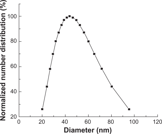
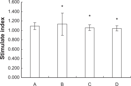
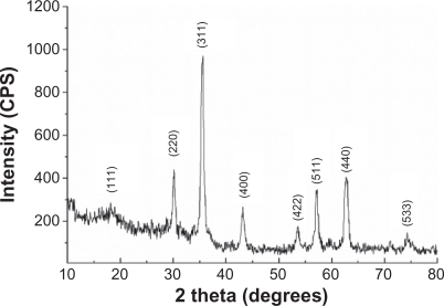
The aim of this article is to study the changes inhibited T lymphocytes and cytokines related to the cellular immunity in ICR (imprinting control region) mice fed with Fe(3)O(4) magnetic nanoparticles (Fe(3)O(4)-MNPs). The Fe(3)O(4)-MNPs were synthesized, and their characteristics such as particle size, zeta potential, and X-ray diffraction patterns were measured and determined. All ICR mice were sacrificed after being exposed to 0, 300, 600, and 1200 mg/kg of Fe(3)O(4)-MNPs by single gastric administration for 14 days. Splenocytes proliferation was indicated with stimulate index by MTT assay; release of cytokines in the serum of ICR mice was detected by enzyme-linked immunosorbent assay, and the phenotypic analyses of T-lymphocyte subsets were performed using flow cytometry. Our results indicated that there were no significant differences in splenocyte proliferation and release of cytokines between exposed and control groups. Furthermore, there was no significant difference in the proportions of T-lymphocyte subsets in the low-dose Fe(3)O(4)-MNPs group when compared to the control group, but the proportions of CD3(+)CD4(+) and CD3(+)CD8(+) T-lymphocyte subsets both in the medium- and high-dose Fe(3)O(4)-MNPs groups were higher than those in the control group. It is concluded that a high dose of Fe(3)O(4)-MNPs, to some extent, could influence in vivo immune function of normal ICR mice.
本文旨在研究 ICR(印记控制区)小鼠喂食 Fe3O4 磁性纳米粒子(Fe3O4-MNPs)后,与细胞免疫相关的抑制性 T 淋巴细胞和细胞因子的变化。合成了 Fe3O4-MNPs,并对其粒径、zeta 电位和 X 射线衍射图谱等特性进行了测量和确定。所有 ICR 小鼠在经口单次给予 0、300、600 和 1200mg/kg Fe3O4-MNPs 14 天后被处死。用 MTT 法检测脾细胞增殖的刺激指数;用酶联免疫吸附试验检测 ICR 小鼠血清中细胞因子的释放情况,并通过流式细胞术进行 T 淋巴细胞亚群的表型分析。结果表明,暴露组与对照组之间的脾细胞增殖和细胞因子释放无显著差异。此外,与对照组相比,低剂量 Fe3O4-MNPs 组 T 淋巴细胞亚群的比例无显著差异,但中、高剂量 Fe3O4-MNPs 组的 CD3+CD4+和 CD3+CD8+T 淋巴细胞亚群比例均高于对照组。结论是,高剂量的 Fe3O4-MNPs 在一定程度上可能会影响正常 ICR 小鼠的体内免疫功能。