National Institute of Nutrition and Seafood Research (NIFES), Bergen, Norway.
PLoS One. 2011;6(6):e20917. doi: 10.1371/journal.pone.0020917. Epub 2011 Jun 7.
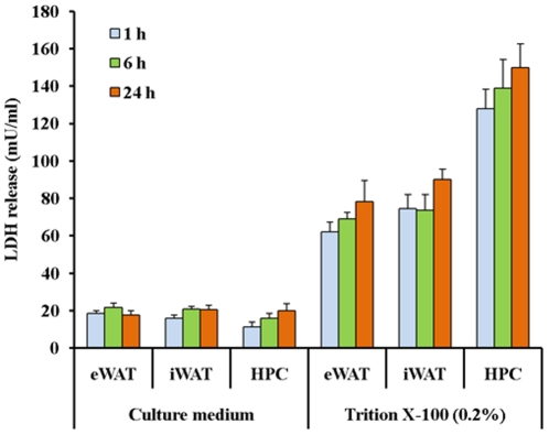
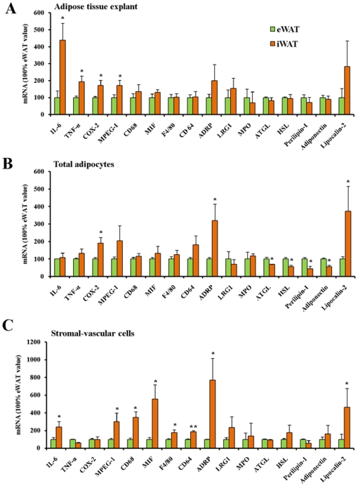
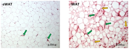
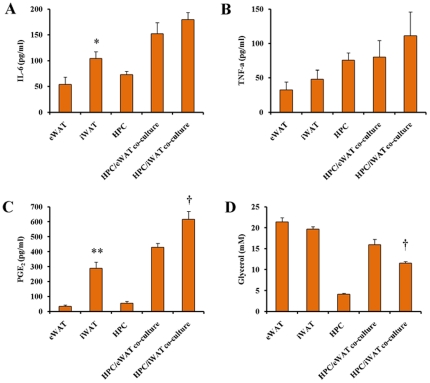
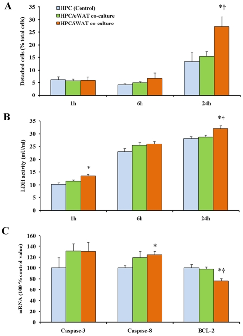
We have developed an in vitro hepatocyte-adipose tissue explant (ATE) co-culture model enabling examination of the effect of visceral and subcutaneous adipose tissues on primary rat hepatocytes. Initial analyses of inflammatory marker genes were performed in fractionated epididymal or inguinal adipose tissues. Expressions of inflammation related genes (IL-6, TNF-α, COX-2) were higher in the inguinal than the epididymal ATE. Similarly, expressions of marker genes of macrophage and monocyte (MPEG-1, CD68, F4/80, CD64) were higher in the stromal vascular fraction (SVF) isolated from inguinal ATE than that from epididymal ATE. However, expressions of lipolysis related genes (ATGL, HSL, perilipin-1) were higher in the epididymal adipocytes than inguinal adipocytes. Moreover, secretion of IL-6 and PGE(2) was higher from inguinal ATEs than from epididymal ATEs. There was a trend that the total levels of IL-6, TNF-α and PGE(2) in the media from inguinal ATEs co-cultured with primary rat hepatocytes were higher than that in the media from epididymal ATEs co-cultured with hepatocytes, although the significant difference was only seen in PGE(2). Lipolysis, measured as glycerol release, was similar in the ATEs isolated from inguinal and epididymal adipose tissues when cultured alone, but the glycerol release was higher in the ATEs isolated from epididymal than from inguinal adipose tissue when co-cultured with hepatocytes. Compared to epididymal ATEs, the ATEs from inguinal adipose tissue elicited a stronger cytotoxic response and higher level of insulin resistance in the co-cultured hepatocytes. In conclusion, our results reveal depot-dependent effects of ATEs on co-cultured primary hepatocytes, which in part may be related to a more pronounced infiltration of stromal vascular cells (SVCs), particularly macrophages, in inguinal adipose tissue resulting in stronger responses in terms of hepatotoxicity and insulin-resistance.
我们开发了一种体外肝细胞-脂肪组织外植体(ATE)共培养模型,可用于研究内脏和皮下脂肪组织对原代大鼠肝细胞的影响。最初在分离的附睾或腹股沟脂肪组织中分析了炎症标志物基因。炎症相关基因(IL-6、TNF-α、COX-2)在腹股沟 ATE 中的表达高于附睾 ATE。同样,从腹股沟 ATE 分离的基质血管部分(SVF)中巨噬细胞和单核细胞(MPEG-1、CD68、F4/80、CD64)的标记基因表达高于附睾 ATE。然而,脂肪分解相关基因(ATGL、HSL、 perilipin-1)在附睾脂肪细胞中的表达高于腹股沟脂肪细胞。此外,IL-6 和 PGE(2)从腹股沟 ATE 的分泌高于附睾 ATE。有一个趋势,即与共培养的原代大鼠肝细胞相比,来自腹股沟 ATE 的培养基中 IL-6、TNF-α 和 PGE(2)的总水平高于来自附睾 ATE 的培养基,尽管仅在 PGE(2)中观察到显著差异。当单独培养时,从腹股沟和附睾脂肪组织分离的 ATE 的脂肪分解(以甘油释放衡量)相似,但当与肝细胞共培养时,从附睾脂肪组织分离的 ATE 的甘油释放高于腹股沟脂肪组织分离的 ATE。与附睾 ATE 相比,腹股沟脂肪组织的 ATE 在共培养的肝细胞中引起更强的细胞毒性反应和更高水平的胰岛素抵抗。总之,我们的结果揭示了 ATE 对共培养原代肝细胞的 depot 依赖性影响,这部分可能与腹股沟脂肪组织中基质血管细胞(SVC)特别是巨噬细胞的浸润更为明显有关,导致在肝毒性和胰岛素抵抗方面的反应更强。