Department of Chemistry & Macromolecules & Interfaces Institute, Virginia Polytechnic Institute and State University, Blacksburg, Virginia 24061, United States.
Mol Pharm. 2011 Oct 3;8(5):1709-19. doi: 10.1021/mp200078n. Epub 2011 Jul 18.
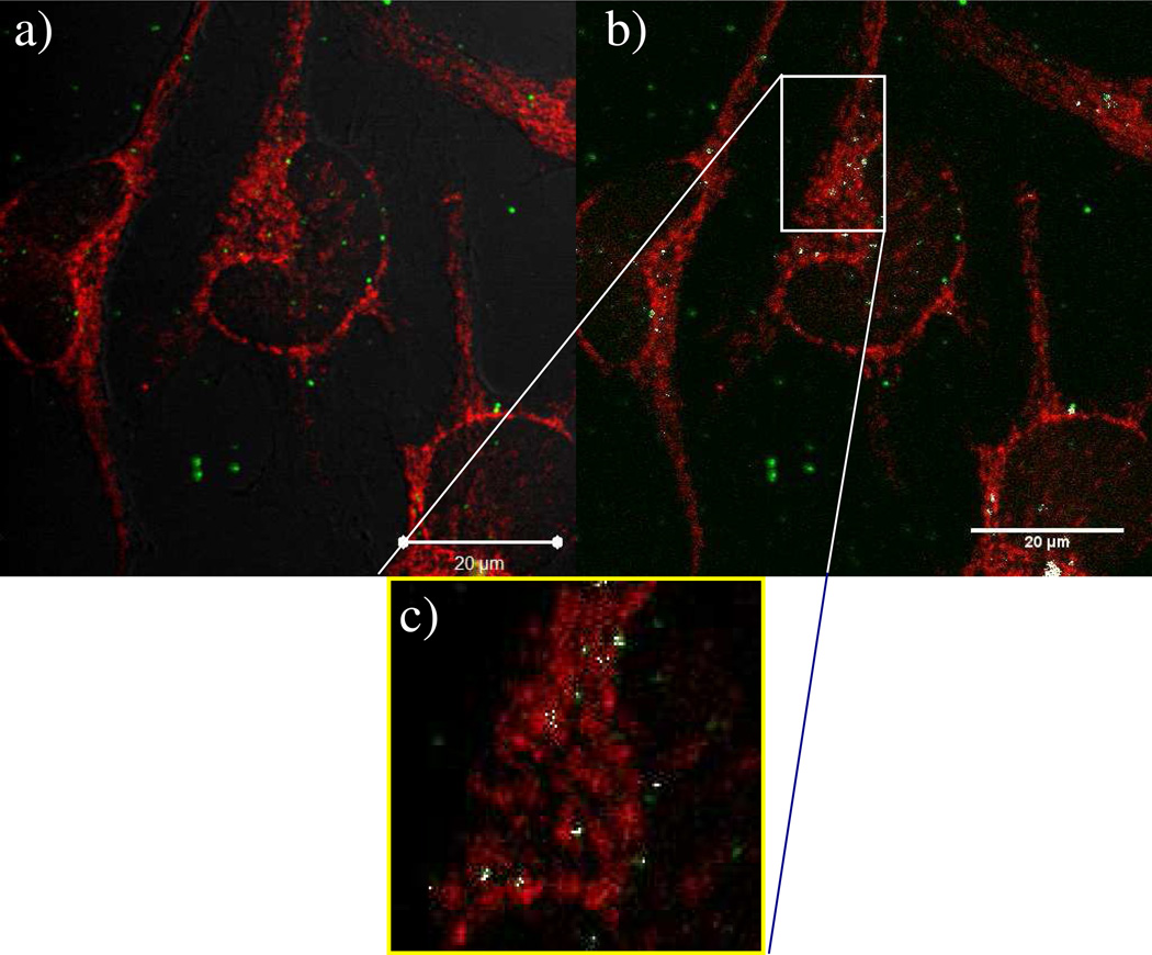
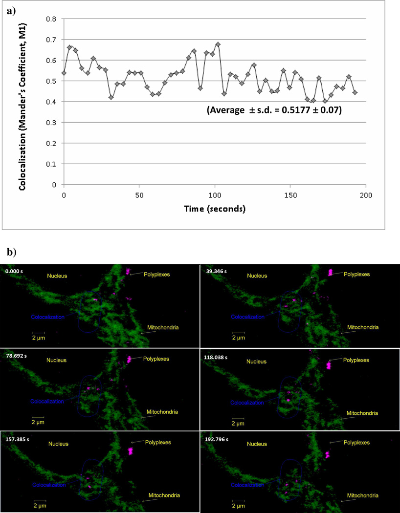
Poly(ethylenimine) (PEI) and PEI-based systems have been widely studied for use as nucleic acid delivery vehicles. However, many of these vehicles display high cytotoxicity, rendering them unfit for therapeutic use. By exploring the mechanisms that cause cytotoxicity, and through understanding structure-function relationships between polymers and intracellular interactions, nucleic acid delivery vehicles with precise intracellular properties can be tailored for specific function. Previous research has shown that PEI is able to depolarize mitochondria, but the exact mechanism as to how depolarization is induced remains elusive and therefore is the focus of the current study. Potential mechanisms for mitochondrial depolarization include direct mitochondrial membrane permeabilization by PEI or PEI polyplexes, activation of the mitochondrial permeability transition pore, and interference with mitochondrial membrane proton pumps, specifically Complex I of the electron transport chain and F(0)F(1)-ATPase. Herein, confocal microscopy and live cell imaging showed that PEI polyplexes do colocalize to some degree with mitochondria early in transfection, and the degree of colocalization increases over time. Cyclosporin a was used to prevent activation of the mitochondrial membrane permeability transition pore, and it was found that early in transfection cyclosporin a was unable to prevent the loss of mitochondrial membrane potential. Further studies done using rotenone and oligomycin to inhibit Complex I of the electron transport chain and F(0)F(1)-ATPase, respectively, indicate that both of these mitochondrial proton pumps are functioning during PEI transfection. Overall, we conclude that direct interaction between polyplexes and mitochondria may be the reason why mitochondrial function is impaired during PEI transfection.
聚乙亚胺(PEI)及其基于 PEI 的系统已被广泛研究用于核酸传递载体。然而,许多这些载体显示出高细胞毒性,使其不适合治疗用途。通过探索导致细胞毒性的机制,以及通过理解聚合物和细胞内相互作用之间的结构-功能关系,可以为特定功能定制具有精确细胞内特性的核酸传递载体。先前的研究表明,PEI 能够使线粒体去极化,但去极化是如何诱导的具体机制仍不清楚,因此是当前研究的重点。线粒体去极化的潜在机制包括 PEI 或 PEI 聚合物直接使线粒体膜通透性增加、激活线粒体通透性转换孔以及干扰线粒体膜质子泵,特别是电子传递链复合物 I 和 F(0)F(1)-ATP 酶。本文通过共聚焦显微镜和活细胞成像显示,PEI 聚合物在转染早期在某种程度上与线粒体共定位,并且共定位程度随时间增加。环孢菌素 A 用于防止线粒体膜通透性转换孔的激活,发现早期转染时环孢菌素 A 无法防止线粒体膜电位的丧失。使用鱼藤酮和寡霉素分别抑制电子传递链复合物 I 和 F(0)F(1)-ATP 酶的进一步研究表明,在 PEI 转染过程中这两种线粒体质子泵都在起作用。总的来说,我们得出结论,聚合物与线粒体之间的直接相互作用可能是 PEI 转染过程中线粒体功能受损的原因。