Department of Periodontology, School of Dental Medicine, University of Bern, Bern, Switzerland.
PLoS One. 2011;6(8):e23375. doi: 10.1371/journal.pone.0023375. Epub 2011 Aug 15.
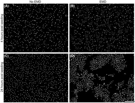
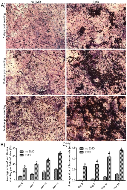
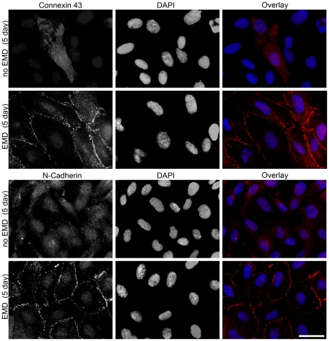
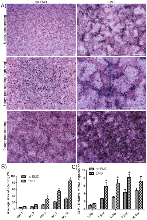
In recent years, enamel matrix derivative (EMD) has garnered much interest in the dental field for its apparent bioactivity that stimulates regeneration of periodontal tissues including periodontal ligament, cementum and alveolar bone. Despite its widespread use, the underlying cellular mechanisms remain unclear and an understanding of its biological interactions could identify new strategies for tissue engineering. Previous in vitro research has demonstrated that EMD promotes premature osteoblast clustering at early time points. The aim of the present study was to evaluate the influence of cell clustering on vital osteoblast cell-cell communication and adhesion molecules, connexin 43 (cx43) and N-cadherin (N-cad) as assessed by immunofluorescence imaging, real-time PCR and Western blot analysis. In addition, differentiation markers of osteoblasts were quantified using alkaline phosphatase, osteocalcin and von Kossa staining. EMD significantly increased the expression of connexin 43 and N-cadherin at early time points ranging from 2 to 5 days. Protein expression was localized to cell membranes when compared to control groups. Alkaline phosphatase activity was also significantly increased on EMD-coated samples at 3, 5 and 7 days post seeding. Interestingly, higher activity was localized to cell cluster regions. There was a 3 fold increase in osteocalcin and bone sialoprotein mRNA levels for osteoblasts cultured on EMD-coated culture dishes. Moreover, EMD significantly increased extracellular mineral deposition in cell clusters as assessed through von Kossa staining at 5, 7, 10 and 14 days post seeding. We conclude that EMD up-regulates the expression of vital osteoblast cell-cell communication and adhesion molecules, which enhances the differentiation and mineralization activity of osteoblasts. These findings provide further support for the clinical evidence that EMD increases the speed and quality of new bone formation in vivo.
近年来,由于其明显的生物活性,牙科学领域对釉基质衍生物 (EMD) 产生了浓厚的兴趣,这种生物活性可刺激牙周组织(包括牙周韧带、牙骨质和牙槽骨)的再生。尽管它被广泛应用,但潜在的细胞机制仍不清楚,对其生物学相互作用的了解可以确定组织工程的新策略。先前的体外研究表明,EMD 可促进成骨细胞在早期阶段过早聚集。本研究旨在评估细胞聚集对重要成骨细胞细胞间通讯和粘附分子的影响,通过免疫荧光成像、实时 PCR 和 Western blot 分析评估连接蛋白 43 (cx43) 和 N-钙粘蛋白 (N-cad)。此外,还通过碱性磷酸酶、骨钙素和 von Kossa 染色定量测定成骨细胞的分化标志物。EMD 显著增加了连接蛋白 43 和 N-钙粘蛋白的表达,在 2 至 5 天的早期时间点。与对照组相比,蛋白表达定位于细胞膜。在接种后 3、5 和 7 天,碱性磷酸酶活性也显著增加。有趣的是,较高的活性定位于细胞簇区域。在 EMD 包被的培养皿中培养的成骨细胞中,骨钙素和骨涎蛋白 mRNA 水平增加了 3 倍。此外,在接种后 5、7、10 和 14 天,通过 von Kossa 染色,EMD 显著增加了细胞簇中外源性矿物质沉积。我们得出结论,EMD 上调了重要的成骨细胞细胞间通讯和粘附分子的表达,从而增强了成骨细胞的分化和矿化活性。这些发现为 EMD 增加体内新骨形成的速度和质量的临床证据提供了进一步支持。