Neurobiological Research Laboratory, Department of Otorhinolaryngology, University of Freiburg, Freiburg, Germany.
PLoS One. 2011;6(8):e23686. doi: 10.1371/journal.pone.0023686. Epub 2011 Aug 22.
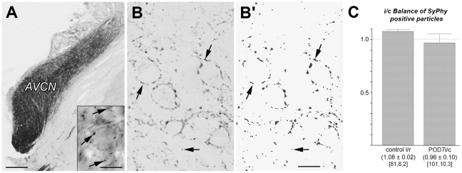
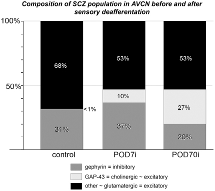
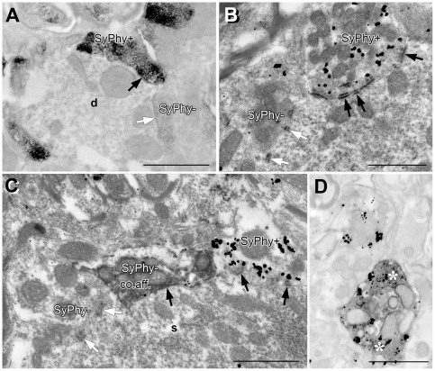
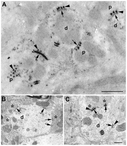
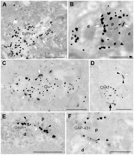
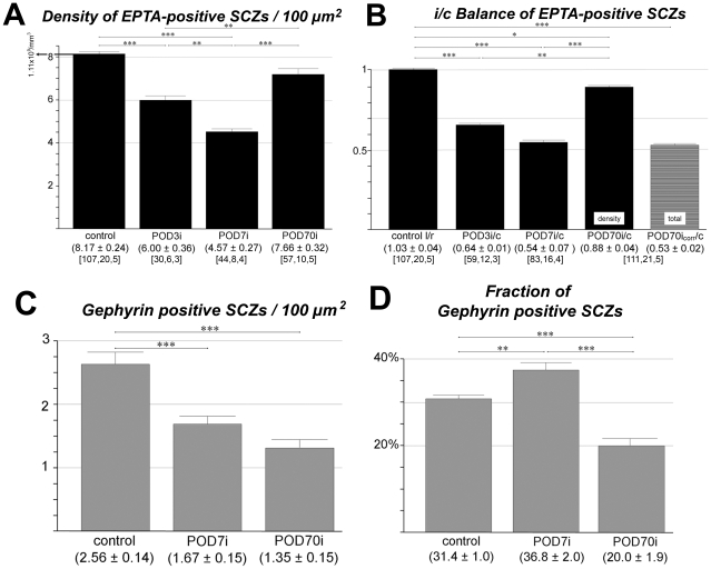
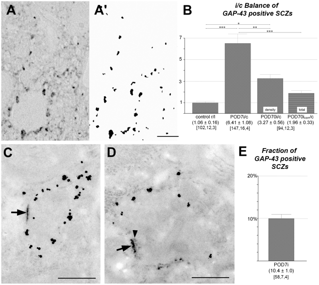
Ablation of a cochlea causes total sensory deafferentation of the cochlear nucleus in the brainstem, providing a model to investigate nervous degeneration and formation of new synaptic contacts in the adult brain. In a quantitative electron microscopical study on the plasticity of the central auditory system of the Wistar rat, we first determined what fraction of the total number of synaptic contact zones (SCZs) in the anteroventral cochlear nucleus (AVCN) is attributable to primary sensory innervation and how many synapses remain after total unilateral cochlear ablation. Second, we attempted to identify the potential for a deafferentation-dependent synaptogenesis. SCZs were ultrastructurally identified before and after deafferentation in tissue treated for ethanolic phosphotungstic acid (EPTA) staining. This was combined with pre-embedding immunocytochemistry for gephyrin identifying inhibitory SCZs, the growth-associated protein GAP-43, glutamate, and choline acetyltransferase. A stereological analysis of EPTA stained sections revealed 1.11±0.09 (S.E.M.)×10(9) SCZs per mm(3) of AVCN tissue. Within 7 days of deafferentation, this number was down by 46%. Excitatory and inhibitory synapses were differentially affected on the side of deafferentation. Excitatory synapses were quickly reduced and then began to increase in number again, necessarily being complemented from sources other than cochlear neurons, while inhibitory synapses were reduced more slowly and continuously. The result was a transient rise of the relative fraction of inhibitory synapses with a decline below original levels thereafter. Synaptogenesis was inferred by the emergence of morphologically immature SCZs that were consistently associated with GAP-43 immunoreactivity. SCZs of this type were estimated to make up a fraction of close to 30% of the total synaptic population present by ten weeks after sensory deafferentation. In conclusion, there appears to be a substantial potential for network reorganization and synaptogenesis in the auditory brainstem after loss of hearing, even in the adult brain.
耳蜗消融会导致脑干耳蜗核的感觉传入完全丧失,为研究成年大脑中的神经退行性变和新突触接触的形成提供了模型。在一项关于威斯塔大鼠中枢听觉系统可塑性的定量电子显微镜研究中,我们首先确定了前腹侧耳蜗核 (AVCN) 中总突触接触区 (SCZ) 的一部分归因于初级感觉传入,以及在完全单侧耳蜗消融后还剩下多少个突触。其次,我们试图确定去传入依赖性突触发生的潜力。在去传入前和去传入后,通过对经乙醇磷钨酸 (EPTA) 染色处理的组织进行超微结构鉴定,确定了 SCZ。这与 GAP-43、谷氨酸和胆碱乙酰转移酶的预包埋免疫细胞化学相结合。EPTA 染色切片的体视学分析显示,AVCN 组织中每立方毫米有 1.11±0.09(S.E.M.)×10(9) 个 SCZ。在去传入的 7 天内,这个数量减少了 46%。传入侧的兴奋性和抑制性突触受到不同的影响。兴奋性突触迅速减少,然后开始再次增加,必然是由耳蜗神经元以外的其他来源补充的,而抑制性突触减少得更慢且持续。结果是抑制性突触的相对分数暂时上升,随后又下降到原始水平以下。突触发生是通过出现形态不成熟的 SCZ 推断出来的,这些 SCZ 始终与 GAP-43 免疫反应性相关。这种类型的 SCZ 估计占感觉传入丧失后 10 周时存在的总突触群体的近 30%。总之,即使在成年大脑中,听觉脑干在听力丧失后似乎也有很大的网络重组和突触发生的潜力。