Department of Radiology, Johns Hopkins University Baltimore, MD, USA.
Front Neurol. 2011 Aug 24;2:54. doi: 10.3389/fneur.2011.00054. eCollection 2011.
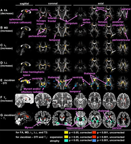
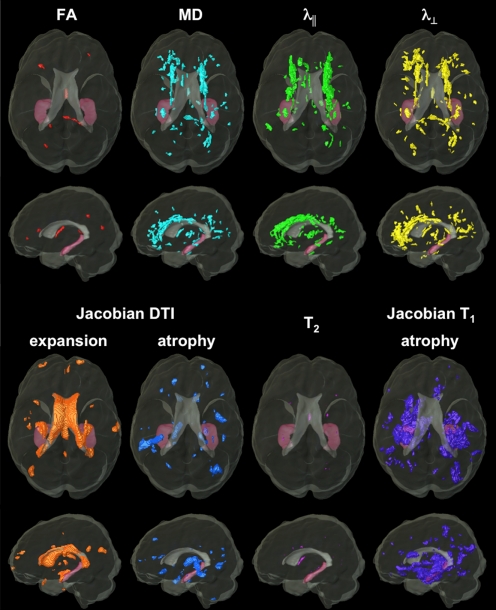
Alterations of the gray and white matter have been identified in Alzheimer's disease (AD) by structural magnetic resonance imaging (MRI) and diffusion tensor imaging (DTI). However, whether the combination of these modalities could increase the diagnostic performance is unknown.
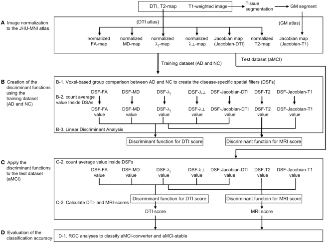
Participants included 19 AD patients, 22 amnestic mild cognitive impairment (aMCI) patients, and 22 cognitively normal elderly (NC). The aMCI group was further divided into an "aMCI-converter" group (converted to AD dementia within 3 years), and an "aMCI-stable" group who did not convert in this time period. A T(1)-weighted image, a T(2) map, and a DTI of each participant were normalized, and voxel-based comparisons between AD and NC groups were performed. Regions-of-interest, which defined the areas with significant differences between AD and NC, were created for each modality and named "disease-specific spatial filters" (DSF). Linear discriminant analysis was used to optimize the combination of multiple MRI measurements extracted by DSF to effectively differentiate AD from NC. The resultant DSF and the discriminant function were applied to the aMCI group to investigate the power to differentiate the aMCI-converters from the aMCI-stable patients.
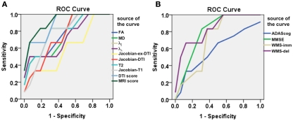
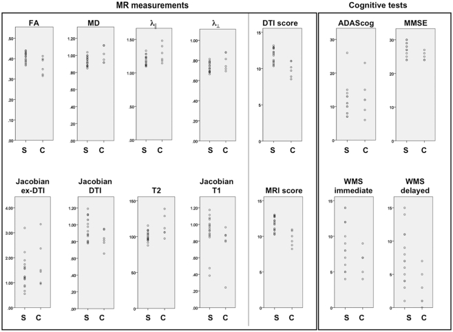
The multi-modal approach with AD-specific filters led to a predictive model with an area under the receiver operating characteristic curve (AUC) of 0.93, in differentiating aMCI-converters from aMCI-stable patients. This AUC was better than that of a single-contrast-based approach, such as T(1)-based morphometry or diffusion anisotropy analysis.
The multi-modal approach has the potential to increase the value of MRI in predicting conversion from aMCI to AD.
结构磁共振成像(MRI)和弥散张量成像(DTI)已在阿尔茨海默病(AD)中发现了灰质和白质的改变。然而,这些模态的组合是否能提高诊断性能尚不清楚。
参与者包括 19 名 AD 患者、22 名遗忘型轻度认知障碍(aMCI)患者和 22 名认知正常的老年人(NC)。aMCI 组进一步分为“aMCI 转化组”(在 3 年内转化为 AD 痴呆)和“aMCI 稳定组”,在此期间未转化。对每位参与者的 T1 加权图像、T2 图谱和 DTI 进行归一化,并对 AD 和 NC 组进行基于体素的比较。为每个模态创建了定义 AD 和 NC 之间存在显著差异的区域的感兴趣区域,并将其命名为“疾病特异性空间滤波器”(DSF)。线性判别分析用于优化通过 DSF 提取的多个 MRI 测量的组合,以有效地区分 AD 与 NC。将所得的 DSF 和判别函数应用于 aMCI 组,以研究其区分 aMCI 转化者与 aMCI 稳定者的能力。
使用 AD 特异性滤波器的多模态方法导致预测模型的受试者工作特征曲线下面积(AUC)为 0.93,可区分 aMCI 转化者与 aMCI 稳定者。该 AUC 优于基于单一对比的方法,如基于 T1 的形态测量或弥散各向异性分析。
多模态方法有可能提高 MRI 在预测从 aMCI 向 AD 转化中的价值。