Univ. Bordeaux, LaBRI, UMR 5800, PICTURA, F-33400, Talence, France.
Bordeaux INP, LaBRI, UMR 5800, PICTURA, F-33405, Talence, France.
Sci Rep. 2019 Sep 25;9(1):13845. doi: 10.1038/s41598-019-49970-9.
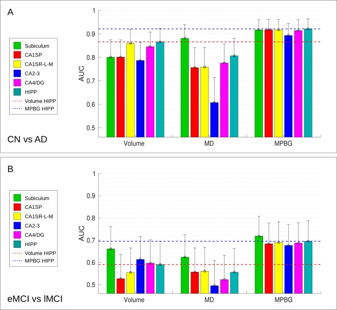
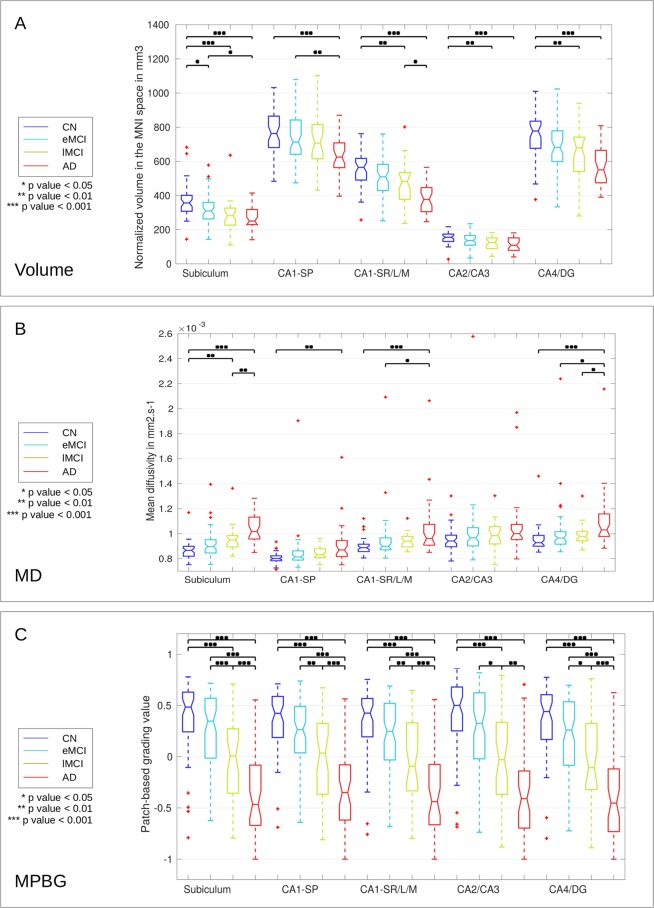
Numerous studies have proposed biomarkers based on magnetic resonance imaging (MRI) to detect and predict the risk of evolution toward Alzheimer's disease (AD). Most of these methods have focused on the hippocampus, which is known to be one of the earliest structures impacted by the disease. To date, patch-based grading approaches provide among the best biomarkers based on the hippocampus. However, this structure is complex and is divided into different subfields, not equally impacted by AD. Former in-vivo imaging studies mainly investigated structural alterations of these subfields using volumetric measurements and microstructural modifications with mean diffusivity measurements. The aim of our work is to improve the current classification performances based on the hippocampus with a new multimodal patch-based framework combining structural and diffusivity MRI. The combination of these two MRI modalities enables the capture of subtle structural and microstructural alterations. Moreover, we propose to study the efficiency of this new framework applied to the hippocampal subfields. To this end, we compare the classification accuracy provided by the different hippocampal subfields using volume, mean diffusivity, and our novel multimodal patch-based grading framework combining structural and diffusion MRI. The experiments conducted in this work show that our new multimodal patch-based method applied to the whole hippocampus provides the most discriminating biomarker for advanced AD detection while our new framework applied into subiculum obtains the best results for AD prediction, improving by two percentage points the accuracy compared to the whole hippocampus.
许多研究基于磁共振成像(MRI)提出了生物标志物,以检测和预测向阿尔茨海默病(AD)发展的风险。这些方法大多集中在海马体上,已知海马体是最早受到疾病影响的结构之一。迄今为止,基于海马体的基于斑块的分级方法提供了其中最好的生物标志物。然而,这个结构很复杂,分为不同的亚区,受 AD 的影响并不均衡。以前的体内成像研究主要使用体积测量和各向异性系数测量的微观结构改变来研究这些亚区的结构改变。我们的工作旨在通过一种新的结合结构和弥散磁共振成像的多模态斑块分级框架来提高基于海马体的现有分类性能。这两种 MRI 模式的结合能够捕捉到细微的结构和微观结构改变。此外,我们还提出了研究该新框架在海马亚区中的应用的效率。为此,我们比较了使用体积、平均扩散系数和我们新的结合结构和弥散 MRI 的多模态斑块分级框架的不同海马亚区提供的分类准确性。本工作中的实验表明,我们的新的基于多模态斑块的方法应用于整个海马体提供了最具区分性的生物标志物,用于高级 AD 检测,而我们的新框架应用于海马旁回则获得了 AD 预测的最佳结果,与整个海马体相比,准确性提高了两个百分点。