• 文献检索
  • 文档翻译
  • 深度研究
  • 学术资讯
  • Suppr Zotero 插件Zotero 插件
  • 邀请有礼
  • 套餐&价格
  • 历史记录
应用&插件
Suppr Zotero 插件Zotero 插件浏览器插件Mac 客户端Windows 客户端微信小程序
定价
高级版会员购买积分包购买API积分包
服务
文献检索文档翻译深度研究API 文档MCP 服务
关于我们
关于 Suppr公司介绍联系我们用户协议隐私条款
关注我们

Suppr 超能文献

核心技术专利:CN118964589B侵权必究
粤ICP备2023148730 号-1Suppr @ 2026

文献检索

告别复杂PubMed语法,用中文像聊天一样搜索,搜遍4000万医学文献。AI智能推荐,让科研检索更轻松。

立即免费搜索

文件翻译

保留排版,准确专业,支持PDF/Word/PPT等文件格式,支持 12+语言互译。

免费翻译文档

深度研究

AI帮你快速写综述,25分钟生成高质量综述,智能提取关键信息,辅助科研写作。

立即免费体验

使用新型蛋白酶激活荧光探针在小鼠体内对组织蛋白酶E阳性肿瘤进行分子成像。

Molecular imaging of Cathepsin E-positive tumors in mice using a novel protease-activatable fluorescent probe.

作者信息

Abd-Elgaliel Wael R, Cruz-Monserrate Zobeida, Logsdon Craig D, Tung Ching-Hsuan

机构信息

Department of Radiology, The Methodist Hospital Research Institute, Weill Cornell Medical College, 6565 Fannin Street, B5-009, Houston, TX 77030, USA.

Department of Cancer Biology, University of Texas, M. D. Anderson Cancer Center, Houston, TX, USA.

出版信息

Mol Biosyst. 2011 Dec;7(12):3207-3213. doi: 10.1039/c1mb05215b. Epub 2011 Sep 20.

DOI:10.1039/c1mb05215b
PMID:21935563
原文链接:https://pmc.ncbi.nlm.nih.gov/articles/PMC4207267/
Abstract

UNLABELLED

The purpose of this study is to demonstrate the ability of imaging Cathepsin E (Cath E) positive tumors in living animals through selective targeting of Cath E proteolytic activity using a sensitive molecular imaging agent.

METHODS

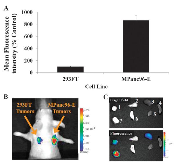
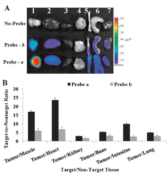
A peptide-based Cath E imaging probe and a control probe were synthesized for this study. Human Cath E-positive cancer cells (MPanc96-E) were implanted subcutaneously in nude mice. Tumor-bearing mice were examined in vivo with near-infrared fluorescence (NIRF) imaging at various time points after intravenous injection of the Cath E sensing imaging probe. Excised organs and tissues of interest were further imaged ex vivo.

RESULTS

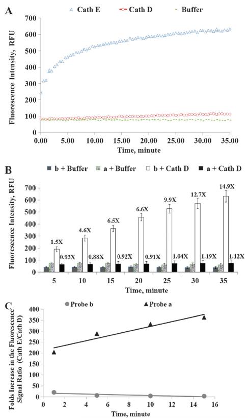
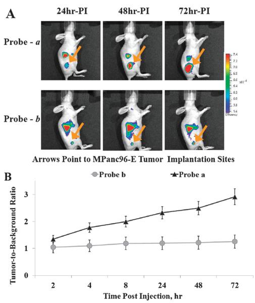
Upon specific Cath E proteolytic activation, the NIRF signal of the imaging probe a was converted from an optically quenched initial state to a highly fluorescent active state. Imaging probe a was able to highlight the Cath E-positive tumors as early as 24 h post injection. Fluorescent signal in tumor was 3-fold higher than background. The confined specificity of imaging probe a to tumor associated Cath E was verified by using control imaging probe b. Both in vivo and ex vivo imaging results confirmed the superior selectivity and sensitivity of imaging probe a in Cath E imaging.

CONCLUSIONS

The small animal studies demonstrated the capability of probe a for imaging Cath E-positive tumors. The developed optical probe could be applied in early diagnostic imaging and guiding subsequent surgical procedure.

摘要

未标记

本研究的目的是通过使用一种灵敏的分子成像剂选择性靶向组织蛋白酶E(Cath E)的蛋白水解活性,来证明在活体动物中对Cath E阳性肿瘤进行成像的能力。

方法

为本研究合成了一种基于肽的Cath E成像探针和一种对照探针。将人Cath E阳性癌细胞(MPanc96-E)皮下植入裸鼠体内。在静脉注射Cath E传感成像探针后的不同时间点,对荷瘤小鼠进行近红外荧光(NIRF)成像体内检查。对切除的感兴趣器官和组织进行进一步的体外成像。

结果

在特定的Cath E蛋白水解激活后,成像探针a的NIRF信号从光学淬灭的初始状态转变为高荧光活性状态。成像探针a在注射后24小时就能突出显示Cath E阳性肿瘤。肿瘤中的荧光信号比背景高3倍。通过使用对照成像探针b验证了成像探针a对肿瘤相关Cath E的局限性特异性。体内和体外成像结果均证实了成像探针a在Cath E成像中的卓越选择性和敏感性。

结论

小动物研究证明了探针a对Cath E阳性肿瘤进行成像的能力。所开发的光学探针可应用于早期诊断成像并指导后续手术操作。

https://cdn.ncbi.nlm.nih.gov/pmc/blobs/9bec/4207267/69963b20f47e/nihms-629087-f0004.jpg
https://cdn.ncbi.nlm.nih.gov/pmc/blobs/9bec/4207267/4b97b03c9286/nihms-629087-f0001.jpg
https://cdn.ncbi.nlm.nih.gov/pmc/blobs/9bec/4207267/e6e200567432/nihms-629087-f0002.jpg
https://cdn.ncbi.nlm.nih.gov/pmc/blobs/9bec/4207267/713e08bb4451/nihms-629087-f0003.jpg
https://cdn.ncbi.nlm.nih.gov/pmc/blobs/9bec/4207267/69963b20f47e/nihms-629087-f0004.jpg

相似文献

1
Molecular imaging of Cathepsin E-positive tumors in mice using a novel protease-activatable fluorescent probe.使用新型蛋白酶激活荧光探针在小鼠体内对组织蛋白酶E阳性肿瘤进行分子成像。
Mol Biosyst. 2011 Dec;7(12):3207-3213. doi: 10.1039/c1mb05215b. Epub 2011 Sep 20.
2
Detection of pancreatic cancer tumours and precursor lesions by cathepsin E activity in mouse models.通过在小鼠模型中组织蛋白酶 E 活性检测胰腺癌肿瘤和前体病变。
Gut. 2012 Sep;61(9):1315-22. doi: 10.1136/gutjnl-2011-300544. Epub 2011 Nov 7.
3
Selective detection of Cathepsin E proteolytic activity.组织蛋白酶E蛋白水解活性的选择性检测
Biochim Biophys Acta. 2010 Sep;1800(9):1002-8. doi: 10.1016/j.bbagen.2010.06.005. Epub 2010 Jun 19.
4
Simultaneous Monitoring of Multi-Enzyme Activity and Concentration in Tumor Using a Triply Labeled Fluorescent In Vivo Imaging Probe.利用三重标记荧光体内成像探针同时监测肿瘤中的多种酶活性和浓度。
Int J Mol Sci. 2020 Apr 27;21(9):3068. doi: 10.3390/ijms21093068.
5
Glucosamine-linked near-infrared fluorescent probes for imaging of solid tumor xenografts.用于实体瘤异种移植成像的葡萄糖胺连接近红外荧光探针。
Mol Imaging Biol. 2012 Aug;14(4):443-51. doi: 10.1007/s11307-011-0520-4.
6
Evaluation of four affibody-based near-infrared fluorescent probes for optical imaging of epidermal growth factor receptor positive tumors.评价四种基于亲和体的近红外荧光探针用于表皮生长因子受体阳性肿瘤的光学成像。
Bioconjug Chem. 2012 Jun 20;23(6):1149-56. doi: 10.1021/bc200596a. Epub 2012 Jun 4.
7
Ex Vivo and In Vivo Noninvasive Imaging of Epidermal Growth Factor Receptor Inhibition on Colon Tumorigenesis Using Activatable Near-Infrared Fluorescent Probes.使用可激活近红外荧光探针在体外和体内对表皮生长因子受体抑制结肠肿瘤发生的无创成像
Mol Imaging. 2017 Jan-Dec;16:1536012117729044. doi: 10.1177/1536012117729044.
8
In vivo near infrared fluorescence (NIRF) intravascular molecular imaging of inflammatory plaque, a multimodal approach to imaging of atherosclerosis.炎症斑块的体内近红外荧光(NIRF)血管内分子成像,一种动脉粥样硬化成像的多模态方法。
J Vis Exp. 2011 Aug 4(54):2257. doi: 10.3791/2257.
9
Multispectral Photoacoustic Imaging of Tumor Protease Activity with a Gold Nanocage-Based Activatable Probe.基于金纳米笼的肿瘤蛋白酶活性的多光谱光声成像的激活型探针。
Mol Imaging Biol. 2018 Dec;20(6):919-929. doi: 10.1007/s11307-018-1203-1.
10
Optical visualization of cathepsin K activity in atherosclerosis with a novel, protease-activatable fluorescence sensor.利用新型蛋白酶可激活荧光传感器对动脉粥样硬化中组织蛋白酶K活性进行光学可视化。
Circulation. 2007 May 1;115(17):2292-8. doi: 10.1161/CIRCULATIONAHA.106.660340. Epub 2007 Apr 9.

引用本文的文献

1
Rising sun or strangled in the cradle? A narrative review of near-infrared fluorescence imaging-guided surgery for pancreatic tumors.初升的太阳还是扼杀在摇篮中?胰腺肿瘤近红外荧光成像引导手术的叙述性综述
Int J Surg. 2024 Dec 1;110(12):7929-7947. doi: 10.1097/JS9.0000000000001676.
2
Potential of uPAR, αvβ6 Integrin, and Tissue Factor as Targets for Molecular Imaging of Oral Squamous Cell Carcinoma: Evaluation of Nine Targets in Primary Tumors and Metastases by Immunohistochemistry.uPAR、αvβ6 整联蛋白和组织因子作为口腔鳞状细胞癌分子成像靶点的潜力:免疫组织化学法评估原发肿瘤和转移灶中的九个靶点。
Int J Mol Sci. 2023 Feb 14;24(4):3853. doi: 10.3390/ijms24043853.
3

本文引用的文献

1
Visible drug delivery by supramolecular nanocarriers directing to single-platformed diagnosis and therapy of pancreatic tumor model.基于超分子纳米载体的可视化药物递释,用于单一平台的胰腺肿瘤模型诊断和治疗。
Cancer Res. 2010 Sep 15;70(18):7031-41. doi: 10.1158/0008-5472.CAN-10-0303. Epub 2010 Aug 4.
2
Selective detection of Cathepsin E proteolytic activity.组织蛋白酶E蛋白水解活性的选择性检测
Biochim Biophys Acta. 2010 Sep;1800(9):1002-8. doi: 10.1016/j.bbagen.2010.06.005. Epub 2010 Jun 19.
3
18F-FDG PET/CT for image-guided and intensity-modulated radiotherapy.
Overview and Future Perspectives on Tumor-Targeted Positron Emission Tomography and Fluorescence Imaging of Pancreatic Cancer in the Era of Neoadjuvant Therapy.
新辅助治疗时代胰腺癌的肿瘤靶向正电子发射断层扫描和荧光成像概述及未来展望
Cancers (Basel). 2021 Dec 2;13(23):6088. doi: 10.3390/cancers13236088.
4
Cathepsin E expression and activity: Role in the detection and treatment of pancreatic cancer.组织蛋白酶 E 的表达和活性:在胰腺癌检测和治疗中的作用。
Pancreatology. 2019 Oct;19(7):951-956. doi: 10.1016/j.pan.2019.09.009. Epub 2019 Sep 20.
5
Advances in Diagnostic and Intraoperative Molecular Imaging of Pancreatic Cancer.胰腺癌诊断与术中分子成像的进展
Pancreas. 2018 Jul;47(6):675-689. doi: 10.1097/MPA.0000000000001075.
6
Imaging the Tumor Microenvironment.肿瘤微环境成像。
Adv Exp Med Biol. 2017;1036:229-257. doi: 10.1007/978-3-319-67577-0_15.
7
Optical imaging probes in oncology.肿瘤学中的光学成像探针。
Oncotarget. 2016 Jul 26;7(30):48753-48787. doi: 10.18632/oncotarget.9066.
8
Monitoring pancreatic carcinogenesis by the molecular imaging of cathepsin E in vivo using confocal laser endomicroscopy.使用共聚焦激光内镜显微镜在体内通过组织蛋白酶E的分子成像监测胰腺癌发生。
PLoS One. 2014 Sep 3;9(9):e106566. doi: 10.1371/journal.pone.0106566. eCollection 2014.
9
Targeting cathepsin E in pancreatic cancer by a small molecule allows in vivo detection.小分子靶向胰腺癌中的组织蛋白酶 E 可实现体内检测。
Neoplasia. 2013 Jul;15(7):684-93. doi: 10.1593/neo.13276.
10
Pancreatic cancer-associated Cathepsin E as a drug activator.胰腺癌相关组织蛋白酶 E 作为一种药物激活剂。
J Control Release. 2013 May 10;167(3):221-7. doi: 10.1016/j.jconrel.2013.02.007. Epub 2013 Feb 26.
用于图像引导和调强放疗的18F-FDG PET/CT
J Nucl Med. 2009 Oct;50(10):1655-65. doi: 10.2967/jnumed.108.055780. Epub 2009 Sep 16.
4
Proteolysis: a biological process adapted in drug delivery, therapy, and imaging.蛋白水解作用:一种适用于药物递送、治疗和成像的生物学过程。
Bioconjug Chem. 2009 Sep;20(9):1683-95. doi: 10.1021/bc800500a.
5
Cathepsins B and D drive hepatic stellate cell proliferation and promote their fibrogenic potential.组织蛋白酶B和D驱动肝星状细胞增殖并增强其纤维化潜能。
Hepatology. 2009 Apr;49(4):1297-307. doi: 10.1002/hep.22753.
6
Highly sensitive detection of early-stage pancreatic cancer by multimodal near-infrared molecular imaging in living mice.通过多模态近红外分子成像在活体小鼠中对早期胰腺癌进行高灵敏度检测。
Int J Cancer. 2008 Nov 1;123(9):2138-47. doi: 10.1002/ijc.23780.
7
Targeted nanoparticles for imaging incipient pancreatic ductal adenocarcinoma.用于早期胰腺导管腺癌成像的靶向纳米颗粒。
PLoS Med. 2008 Apr 15;5(4):e85. doi: 10.1371/journal.pmed.0050085.
8
Gemcitabine chemotherapy and single-fraction stereotactic body radiotherapy for locally advanced pancreatic cancer.吉西他滨化疗联合单次分割立体定向体部放疗治疗局部晚期胰腺癌。
Int J Radiat Oncol Biol Phys. 2008 Nov 1;72(3):678-86. doi: 10.1016/j.ijrobp.2008.01.051. Epub 2008 Apr 18.
9
Cathepsin E: a mini review.组织蛋白酶E:一篇小型综述。
Biochem Biophys Res Commun. 2008 Mar 14;367(3):517-22. doi: 10.1016/j.bbrc.2007.12.163. Epub 2008 Jan 4.
10
Optical imaging: current applications and future directions.光学成像:当前应用与未来方向。
J Nucl Med. 2008 Jan;49(1):1-4. doi: 10.2967/jnumed.107.045799. Epub 2007 Dec 12.