• 文献检索
  • 文档翻译
  • 深度研究
  • 学术资讯
  • Suppr Zotero 插件Zotero 插件
  • 邀请有礼
  • 套餐&价格
  • 历史记录
应用&插件
Suppr Zotero 插件Zotero 插件浏览器插件Mac 客户端Windows 客户端微信小程序
定价
高级版会员购买积分包购买API积分包
服务
文献检索文档翻译深度研究API 文档MCP 服务
关于我们
关于 Suppr公司介绍联系我们用户协议隐私条款
关注我们

Suppr 超能文献

核心技术专利:CN118964589B侵权必究
粤ICP备2023148730 号-1Suppr @ 2026

文献检索

告别复杂PubMed语法,用中文像聊天一样搜索,搜遍4000万医学文献。AI智能推荐,让科研检索更轻松。

立即免费搜索

文件翻译

保留排版,准确专业,支持PDF/Word/PPT等文件格式,支持 12+语言互译。

免费翻译文档

深度研究

AI帮你快速写综述,25分钟生成高质量综述,智能提取关键信息,辅助科研写作。

立即免费体验

高分辨率光学相干断层扫描视网膜成像:一个说明潜力和局限性的病例系列

High-resolution optical coherence tomography retinal imaging: a case series illustrating potential and limitations.

作者信息

Puzyeyeva Olena, Lam Wai Ching, Flanagan John G, Brent Michael H, Devenyi Robert G, Mandelcorn Mark S, Wong Tien, Hudson Christopher

机构信息

Retina Research Group, Department of Ophthalmology and Vision Sciences, University of Toronto, Toronto, ON, M5T 2S8, Canada.

出版信息

J Ophthalmol. 2011;2011:764183. doi: 10.1155/2011/764183. Epub 2011 Sep 29.

DOI:10.1155/2011/764183
PMID:21969910
原文链接:https://pmc.ncbi.nlm.nih.gov/articles/PMC3182576/
Abstract

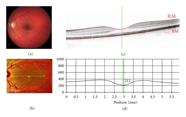
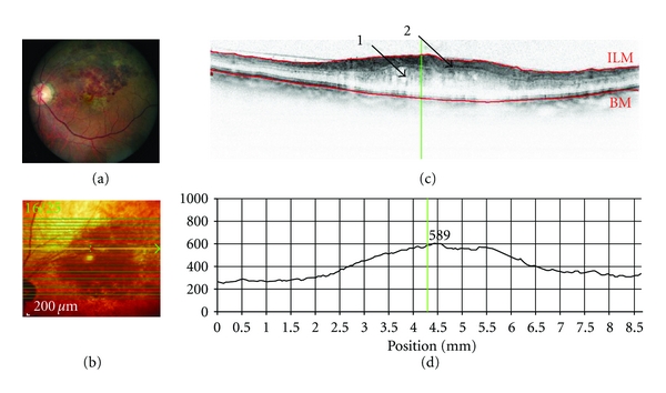
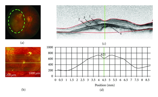
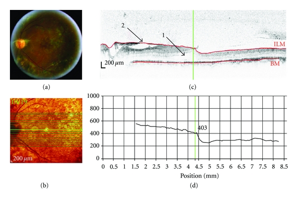
Purpose. To present a series of retinal disease cases that were imaged by spectral domain optical coherence tomography (SD-OCT) in order to illustrate the potential and limitations of this new imaging modality. Methods. The series comprised four selected cases (one case each) of age-related macular degeneration (ARMD), diabetic retinopathy (DR), central retinal artery occlusion (CRAO), and branch retinal vein occlusion (BRVO). Patients were imaged using the Heidelberg Spectralis (Heidelberg Engineering, Germany) in SD-OCT mode. Patients also underwent digital fundus photography and clinical assessment. Results. SD-OCT imaging of a case of age-related macular degeneration revealed a subfoveal choroidal neovascular membrane with detachment of the retinal pigment epithelium (RPE) and neurosensory retina. Using SD-OCT, the cases of DR and BRVO both exhibited macular edema with cystoid spaces visible in the outer retina. Conclusions. The ability of SD-OCT to clearly and objectively elucidate subtle morphological changes within the retinal layers provides information that can be used to formulate diagnoses with greater confidence.

摘要

目的。展示一系列通过光谱域光学相干断层扫描(SD - OCT)成像的视网膜疾病病例,以说明这种新成像方式的潜力和局限性。方法。该系列包括年龄相关性黄斑变性(ARMD)、糖尿病性视网膜病变(DR)、视网膜中央动脉阻塞(CRAO)和视网膜分支静脉阻塞(BRVO)各一例的四个选定病例。患者使用德国海德堡工程公司的海德堡Spectralis进行SD - OCT模式成像。患者还接受了数字眼底摄影和临床评估。结果。年龄相关性黄斑变性一例的SD - OCT成像显示黄斑下脉络膜新生血管膜伴视网膜色素上皮(RPE)和神经感觉视网膜脱离。使用SD - OCT,DR和BRVO病例均表现为黄斑水肿,在外层视网膜可见囊样间隙。结论。SD - OCT清晰、客观地阐明视网膜各层细微形态变化的能力提供了可用于更有信心地做出诊断的信息。

https://cdn.ncbi.nlm.nih.gov/pmc/blobs/0383/3182576/38b963bf7af7/JOP2011-764183.005.jpg
https://cdn.ncbi.nlm.nih.gov/pmc/blobs/0383/3182576/49820f8ee483/JOP2011-764183.001.jpg
https://cdn.ncbi.nlm.nih.gov/pmc/blobs/0383/3182576/9a727541f018/JOP2011-764183.002.jpg
https://cdn.ncbi.nlm.nih.gov/pmc/blobs/0383/3182576/fadccaf22c45/JOP2011-764183.003.jpg
https://cdn.ncbi.nlm.nih.gov/pmc/blobs/0383/3182576/1763c0f8583e/JOP2011-764183.004.jpg
https://cdn.ncbi.nlm.nih.gov/pmc/blobs/0383/3182576/38b963bf7af7/JOP2011-764183.005.jpg

相似文献

1
High-resolution optical coherence tomography retinal imaging: a case series illustrating potential and limitations.高分辨率光学相干断层扫描视网膜成像:一个说明潜力和局限性的病例系列
J Ophthalmol. 2011;2011:764183. doi: 10.1155/2011/764183. Epub 2011 Sep 29.
2
SD-OCT pattern of retinal venous occlusion with cystoid macular edema treated with Ozurdex®.玻璃体内注射Ozurdex®治疗视网膜静脉阻塞合并黄斑囊样水肿的频域光学相干断层扫描图像特征
Eur J Ophthalmol. 2011 Sep-Oct;21(5):631-6. doi: 10.5301/EJO.2011.7428.
3
Retinal and choroidal changes and visual outcome in central retinal artery occlusion: an optical coherence tomography study.视网膜中央动脉阻塞时的视网膜和脉络膜变化及视觉预后:一项光学相干断层扫描研究
Am J Ophthalmol. 2015 Apr;159(4):667-76. doi: 10.1016/j.ajo.2015.01.001. Epub 2015 Jan 9.
4
Predicting visual outcomes for macular disease using optical coherence tomography.使用光学相干断层扫描预测黄斑疾病的视觉预后。
Saudi J Ophthalmol. 2011 Apr;25(2):145-58. doi: 10.1016/j.sjopt.2011.01.003. Epub 2011 Jan 26.
5
Combined confocal scanning laser ophthalmoscopy and spectral-domain optical coherence tomography imaging of reticular drusen associated with age-related macular degeneration.联合共焦扫描激光检眼镜和频域光相干断层扫描成像在年龄相关性黄斑变性相关的网状玻璃膜疣的应用。
Ophthalmology. 2010 Jun;117(6):1169-76. doi: 10.1016/j.ophtha.2009.10.044. Epub 2010 Feb 16.
6
In-vivo mapping of drusen by fundus autofluorescence and spectral-domain optical coherence tomography imaging.通过眼底自发荧光和光谱域光学相干断层扫描成像对玻璃膜疣进行体内成像。
Graefes Arch Clin Exp Ophthalmol. 2016 Jan;254(1):59-67. doi: 10.1007/s00417-015-3012-4. Epub 2015 Apr 24.
7
[Spectral domain OCT in eyes with retinal artery occlusion].视网膜动脉阻塞患者眼部的频域光学相干断层扫描
J Fr Ophtalmol. 2012 Oct;35(8):606-13. doi: 10.1016/j.jfo.2012.04.008. Epub 2012 Jul 21.
8
[Pathophysiology of macular diseases--morphology and function].黄斑疾病的病理生理学——形态与功能
Nippon Ganka Gakkai Zasshi. 2011 Mar;115(3):238-74; discussion 275.
9
Relationship between fluorescein pooling and optical coherence tomographic reflectivity of cystoid spaces in diabetic macular edema.糖尿病性黄斑水肿中囊样间隙的荧光素蓄积与光相干断层扫描反射率的关系。
Ophthalmology. 2012 May;119(5):1047-55. doi: 10.1016/j.ophtha.2011.10.030. Epub 2012 Feb 11.
10
A systematic comparison of spectral-domain optical coherence tomography and fundus autofluorescence in patients with geographic atrophy.光谱域光学相干断层扫描与眼底自发荧光在地图样萎缩患者中的系统比较。
Ophthalmology. 2011 Sep;118(9):1844-51. doi: 10.1016/j.ophtha.2011.01.043. Epub 2011 Apr 15.

引用本文的文献

1
Generalizable Deep Learning for the Detection of Incomplete and Complete Retinal Pigment Epithelium and Outer Retinal Atrophy: A MACUSTAR Report.用于检测不完全和完全视网膜色素上皮和外层视网膜萎缩的可推广深度学习:MACUSTAR 报告。
Transl Vis Sci Technol. 2024 Sep 3;13(9):11. doi: 10.1167/tvst.13.9.11.
2
Detectability of Retinal Diffusion Restriction in Central Retinal Artery Occlusion is Linked to Inner Retinal Layer Thickness.视网膜中央动脉阻塞中视网膜弥散受限的可检测性与内视网膜层厚度有关。
Clin Neuroradiol. 2022 Dec;32(4):1037-1044. doi: 10.1007/s00062-022-01168-9. Epub 2022 May 3.
3
Semi-Automated Quantification of Retinal and Choroidal Biomarkers in Retinal Vascular Diseases: Agreement of Spectral-Domain Optical Coherence Tomography with and without Enhanced Depth Imaging Mode.

本文引用的文献

1
Clinical evaluation of simultaneous confocal scanning laser ophthalmoscopy imaging combined with high-resolution, spectral-domain optical coherence tomography.共焦激光扫描检眼镜成像联合高分辨率、频域光相干断层扫描的临床评估
Acta Ophthalmol. 2010 Dec;88(8):842-9. doi: 10.1111/j.1755-3768.2009.01602.x.
2
Spectral domain optical coherence tomography imaging of retinal diseases in Singapore.新加坡视网膜疾病的光谱域光学相干断层扫描成像
Ophthalmic Surg Lasers Imaging. 2009 May-Jun;40(3):336-41. doi: 10.3928/15428877-20090430-23.
3
Selected clinical comparisons of spectral domain and time domain optical coherence tomography.
视网膜血管疾病中视网膜和脉络膜生物标志物的半自动定量分析:谱域光学相干断层扫描在有无增强深度成像模式下的一致性
Diagnostics (Basel). 2022 Jan 27;12(2):333. doi: 10.3390/diagnostics12020333.
4
Circumpapillary Retinal Nerve Fiber Layer OCT Imaging in a Parkinson's Disease Cohort-A Multidisciplinary Approach in a Clinical Research Hospital.帕金森病队列中的视乳头周围视网膜神经纤维层光学相干断层扫描成像——临床研究医院的多学科方法
J Pers Med. 2022 Jan 9;12(1):80. doi: 10.3390/jpm12010080.
5
Changes of Peripapillary Retinal Nerve Fiber Layer in Childhood Glaucoma: A Systematic Review and Meta-Analysis.儿童青光眼患者视乳头周围视网膜神经纤维层的变化:一项系统评价与Meta分析
Front Med (Lausanne). 2021 Oct 11;8:740152. doi: 10.3389/fmed.2021.740152. eCollection 2021.
6
Management of ocular surface squamous neoplasia: Bowman Club Lecture 2021.眼表鳞状上皮肿瘤的管理:2021年鲍曼俱乐部讲座
BMJ Open Ophthalmol. 2021 Jul 27;6(1):e000842. doi: 10.1136/bmjophth-2021-000842. eCollection 2021.
7
In vivo Structural Assessments of Ocular Disease in Rodent Models using Optical Coherence Tomography.使用光学相干断层扫描对啮齿动物模型眼部疾病进行的体内结构评估
J Vis Exp. 2020 Jul 24(161). doi: 10.3791/61588.
8
Intraretinal Fluid Pattern Characterization in Optical Coherence Tomography Images.视网膜内液模式特征在光学相干断层扫描图像中的分析。
Sensors (Basel). 2020 Apr 3;20(7):2004. doi: 10.3390/s20072004.
9
Artefact-free topography based scleral-asymmetry.基于无伪影的巩膜不对称性的 topography。
PLoS One. 2019 Jul 26;14(7):e0219789. doi: 10.1371/journal.pone.0219789. eCollection 2019.
10
Artery/Vein Vessel Tree Identification in Near-Infrared Reflectance Retinographies.近红外反射视网膜图像中的动脉/静脉血管树识别。
J Digit Imaging. 2019 Dec;32(6):947-962. doi: 10.1007/s10278-019-00235-x.
Ophthalmic Surg Lasers Imaging. 2009 May-Jun;40(3):325-8. doi: 10.3928/15428877-20090430-20.
4
Clinical use and research applications of Heidelberg retinal angiography and spectral-domain optical coherence tomography - a review.海德堡视网膜血管造影和频域光学相干断层扫描的临床应用与研究进展——综述
Clin Exp Ophthalmol. 2009 Jan;37(1):130-43. doi: 10.1111/j.1442-9071.2009.02017.x.
5
Cellular and functional optical coherence tomography of the human retina: the Cogan lecture.人类视网膜的细胞与功能光学相干断层扫描:科根讲座
Invest Ophthalmol Vis Sci. 2007 Dec;48(12):5339-51. doi: 10.1167/iovs.07-0895.
6
State-of-the-art retinal optical coherence tomography.视网膜光学相干断层扫描技术的最新进展。
Prog Retin Eye Res. 2008 Jan;27(1):45-88. doi: 10.1016/j.preteyeres.2007.07.005. Epub 2007 Aug 11.
7
Improved signal-to-noise ratio in spectral-domain compared with time-domain optical coherence tomography.与时域光学相干断层扫描相比,光谱域中的信噪比得到了改善。
Opt Lett. 2003 Nov 1;28(21):2067-9. doi: 10.1364/ol.28.002067.
8
In vivo human retinal imaging by Fourier domain optical coherence tomography.傅里叶域光学相干断层扫描对人体视网膜进行活体成像。
J Biomed Opt. 2002 Jul;7(3):457-63. doi: 10.1117/1.1482379.