Helmy Ashraf A, Hamad Mostafa A, Aly Ahmed M, Sherif Tahra, Hashem Mostafa, El-Sers Dalia Ah, Semieka Mohammad
Department of General Surgery, Faculty of Medicine, Assiut University, Assiut, Egypt.
Ann Surg Innov Res. 2011 Oct 10;5:8. doi: 10.1186/1750-1164-5-8.
Biliary tract reconstruction continues to be a challenging surgical problem. Multiple experimental attempts have been reported to reconstruct biliary defects with different materials and variable outcome. Our aim was to evaluate a new method for biliary reconstruction using an isolated pedicled gastric tube in a live animal trial and also to present the first clinical case.
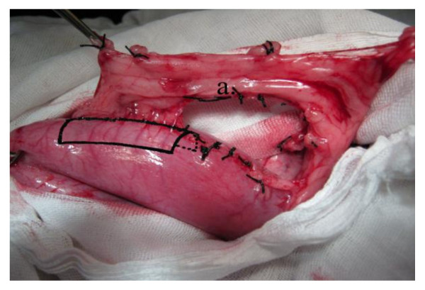
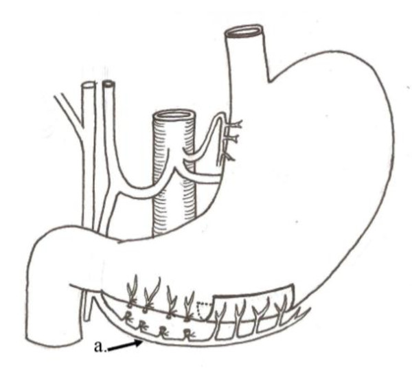
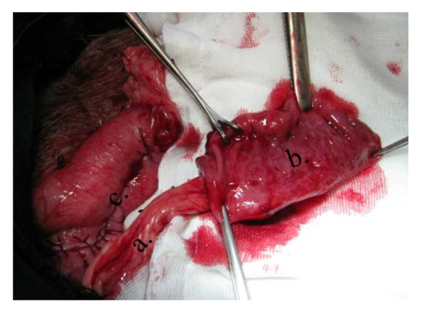
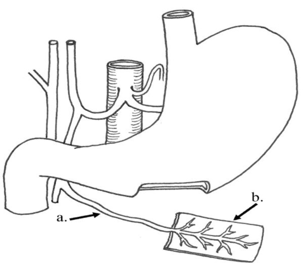
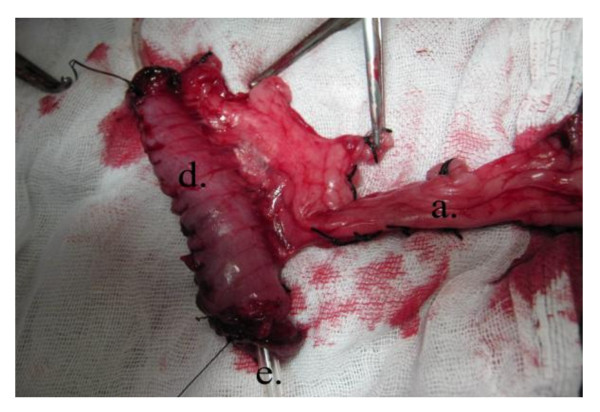
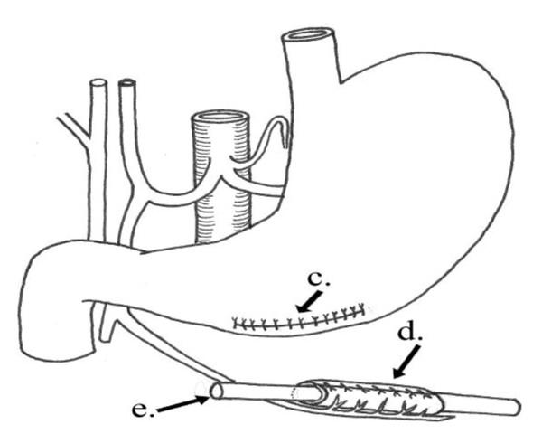
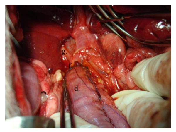
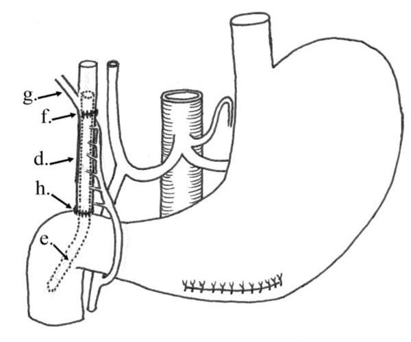
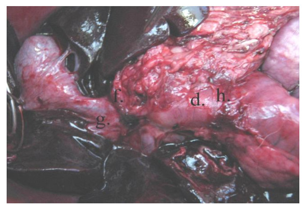
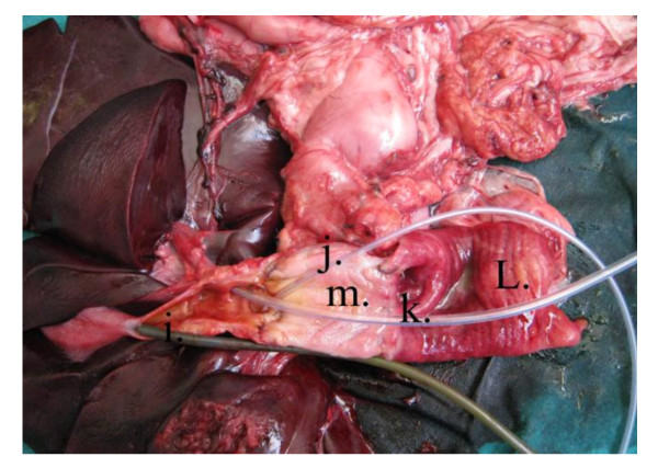
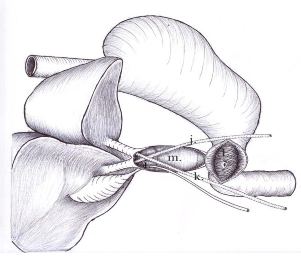
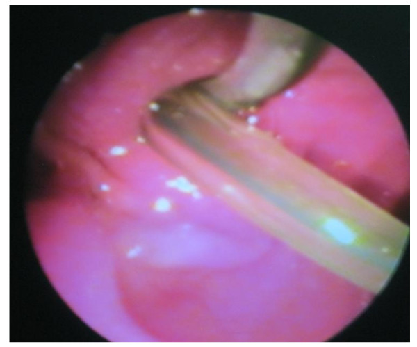
Seven mongrel dogs underwent biliary reconstruction using gastric tube harvested, completely separated from the greater curvature, and based on a vascularized pedicle with the right gastroepiploic vessels. The tube was interposed between the common bile duct (CBD) and the duodenum. Postoperative mortality, morbidity, liver functions, gross and microscopic histological picture were assessed. The first clinical case was also presented where, in a patient with post-cholecystectomy biliary injury, an isolated pedicled gastric tube was interposed between the proximal and distal ends of the CBD.
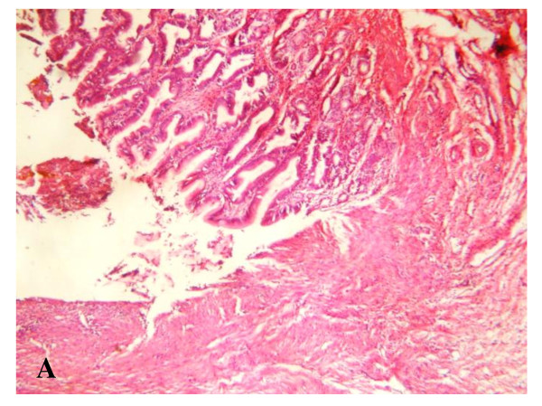
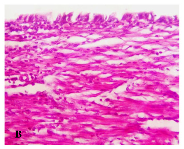
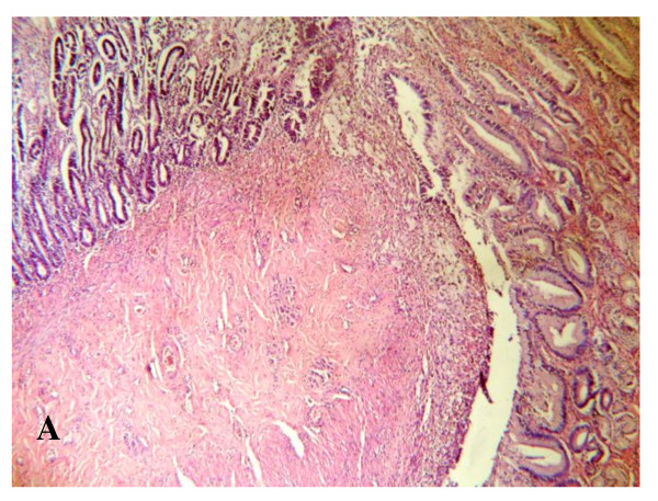
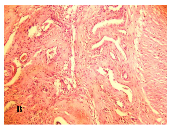
One dog did not recover from anesthesia and another one died postoperatively from septic peritonitis. Five dogs survived the procedure and showed uneventful course and no cholestasis. The mean anastomotic circumference was 4.8 mm (range 4-6) for CBD anastomosis and 6.2 mm (range 5-7) for duodenal anastomosis. Histologically, anastomotic sites showed good evidence of healing. In the first clinical case, the patient showed clinical and biochemical improvement. Endoscopic retrograde cholangiography was feasible and assured patent biliary anastomoses.
In mongrel dogs, biliary reconstruction using pedicled gastric tube interposition between CBD and duodenum is feasible with satisfactory clinical results, anastomotic circumference and histological evidence of healing. The technique is also feasible in human and seems to be promising.
胆道重建仍然是一个具有挑战性的外科问题。据报道,已经进行了多次实验尝试,使用不同材料重建胆道缺损,结果各异。我们的目的是在活体动物试验中评估一种使用孤立带蒂胃管进行胆道重建的新方法,并展示首例临床病例。
七只杂种犬接受了胆道重建,使用从大弯处完全分离并基于带血管蒂的右胃网膜血管的胃管。该胃管置于胆总管(CBD)和十二指肠之间。评估术后死亡率、发病率、肝功能、大体和显微镜下组织学情况。还展示了首例临床病例,在一名胆囊切除术后胆道损伤的患者中,将一根孤立的带蒂胃管置于CBD的近端和远端之间。
一只犬未从麻醉中恢复,另一只犬术后死于感染性腹膜炎。五只犬手术存活,病程平稳,无胆汁淤积。CBD吻合口的平均吻合周长为4.8毫米(范围4 - 6),十二指肠吻合口为6.2毫米(范围5 - 7)。组织学上,吻合部位显示出良好的愈合迹象。在首例临床病例中,患者临床和生化指标均有改善。内镜逆行胆管造影可行,确保了胆道吻合口通畅。
在杂种犬中,将带蒂胃管置于CBD和十二指肠之间进行胆道重建是可行的,临床结果、吻合周长和愈合的组织学证据均令人满意。该技术在人类中也是可行的,似乎很有前景。