• 文献检索
  • 文档翻译
  • 深度研究
  • 学术资讯
  • Suppr Zotero 插件Zotero 插件
  • 邀请有礼
  • 套餐&价格
  • 历史记录
应用&插件
Suppr Zotero 插件Zotero 插件浏览器插件Mac 客户端Windows 客户端微信小程序
定价
高级版会员购买积分包购买API积分包
服务
文献检索文档翻译深度研究API 文档MCP 服务
关于我们
关于 Suppr公司介绍联系我们用户协议隐私条款
关注我们

Suppr 超能文献

核心技术专利:CN118964589B侵权必究
粤ICP备2023148730 号-1Suppr @ 2026

文献检索

告别复杂PubMed语法,用中文像聊天一样搜索,搜遍4000万医学文献。AI智能推荐,让科研检索更轻松。

立即免费搜索

文件翻译

保留排版,准确专业,支持PDF/Word/PPT等文件格式,支持 12+语言互译。

免费翻译文档

深度研究

AI帮你快速写综述,25分钟生成高质量综述,智能提取关键信息,辅助科研写作。

立即免费体验

用于卵巢组织表征的集成光学相干断层扫描、超声和光声成像

Integrated optical coherence tomography, ultrasound and photoacoustic imaging for ovarian tissue characterization.

作者信息

Yang Yi, Li Xiang, Wang Tianheng, Kumavor Patrick D, Aguirre Andres, Shung Kirk K, Zhou Qifa, Sanders Melinda, Brewer Molly, Zhu Quing

出版信息

Biomed Opt Express. 2011 Sep 1;2(9):2551-61. doi: 10.1364/BOE.2.002551. Epub 2011 Aug 5.

DOI:10.1364/BOE.2.002551
PMID:21991547
原文链接:https://pmc.ncbi.nlm.nih.gov/articles/PMC3184864/
Abstract

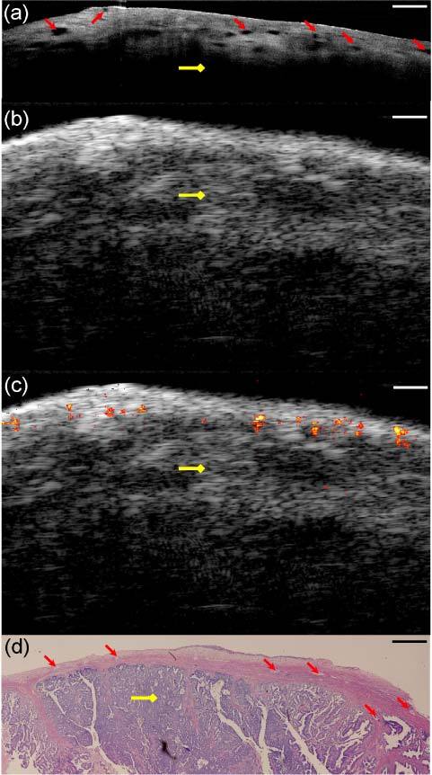
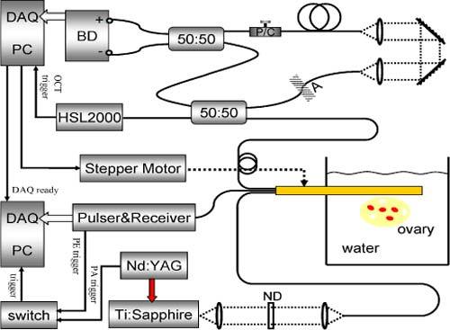
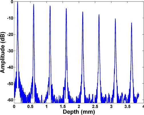
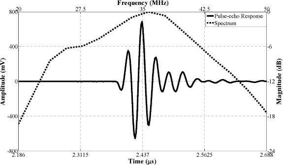
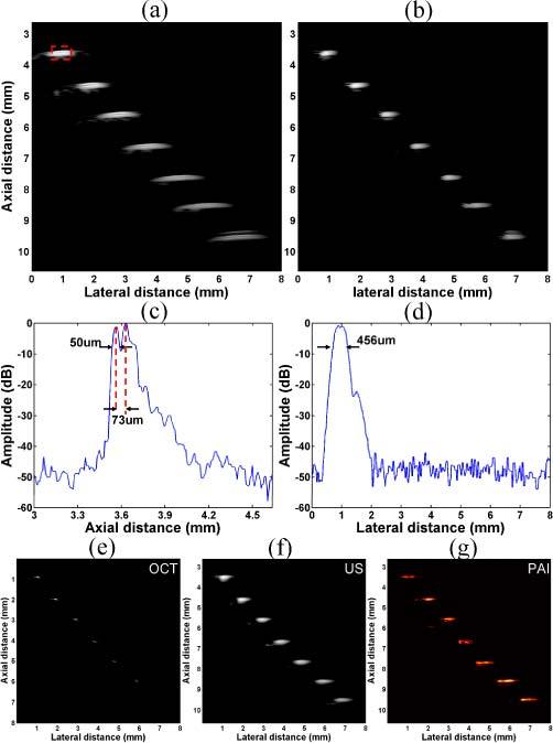
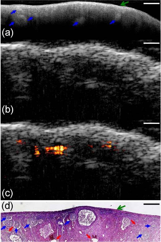
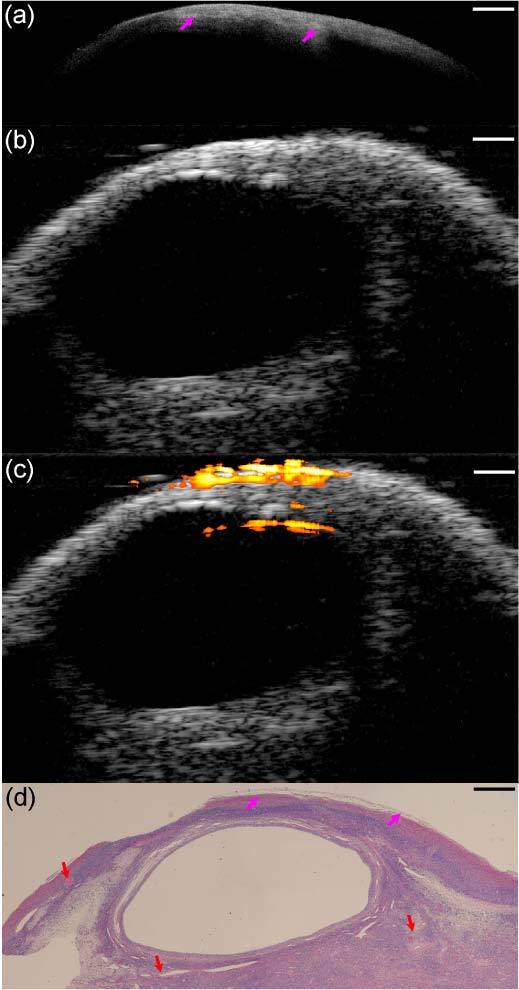
Ovarian cancer has the lowest survival rate of the gynecologic cancers because it is predominantly diagnosed in Stages III or IV due to the lack of reliable symptoms, as well as the lack of efficacious screening techniques. Detection before the malignancy spreads or at the early stage would greatly improve the survival and benefit patient health. In this report, we present an integrated optical coherence tomography (OCT), ultrasound (US) and photoacoustic imaging (PAI) prototype endoscopy system for ovarian tissue characterization. The overall diameter of the prototype endoscope is 5 mm which is suitable for insertion through a standard 5-12.5mm endoscopic laparoscopic port during minimally invasive surgery. It consists of a ball-lensed OCT sample arm probe, a multimode fiber having the output end polished at 45 degree angle so as to deliver the light perpendicularly for PAI, and a high frequency ultrasound transducer with 35MHz center frequency. System characterizations of OCT, US and PAI are presented. In addition, results obtained from ex vivo porcine and human ovarian tissues are presented. The optical absorption contrast provided by PAI, the high resolution subsurface morphology provided by OCT, and the deeper tissue structure imaged by US demonstrate the synergy of the combined endoscopy and the superior performance of this hybrid device over each modality alone in ovarian tissue characterization.

摘要

卵巢癌在妇科癌症中的生存率最低,这是因为其主要在III期或IV期才被诊断出来,原因在于缺乏可靠的症状以及有效的筛查技术。在恶性肿瘤扩散之前或早期进行检测将极大地提高生存率并有益于患者健康。在本报告中,我们展示了一种用于卵巢组织表征的集成光学相干断层扫描(OCT)、超声(US)和光声成像(PAI)的原型内窥镜系统。该原型内窥镜的整体直径为5毫米,适合在微创手术期间通过标准的5 - 12.5毫米内窥镜腹腔镜端口插入。它由一个球透镜OCT样品臂探头、一个输出端以45度角抛光以便垂直传输光用于PAI的多模光纤以及一个中心频率为35MHz的高频超声换能器组成。展示了OCT、US和PAI的系统特性。此外,还展示了从离体猪和人卵巢组织获得的结果。PAI提供的光吸收对比度、OCT提供的高分辨率亚表面形态以及US成像的更深组织结构证明了组合内窥镜的协同作用以及该混合设备在卵巢组织表征方面相对于单一模态的卓越性能。

https://cdn.ncbi.nlm.nih.gov/pmc/blobs/4b3b/3184864/cd9c1c9ca9a8/boe-2-9-2551-g008.jpg
https://cdn.ncbi.nlm.nih.gov/pmc/blobs/4b3b/3184864/eb21d2776106/boe-2-9-2551-g001.jpg
https://cdn.ncbi.nlm.nih.gov/pmc/blobs/4b3b/3184864/9d114b1acab4/boe-2-9-2551-g002.jpg
https://cdn.ncbi.nlm.nih.gov/pmc/blobs/4b3b/3184864/3e75c1a49b94/boe-2-9-2551-g003.jpg
https://cdn.ncbi.nlm.nih.gov/pmc/blobs/4b3b/3184864/4629302be2cc/boe-2-9-2551-g004.jpg
https://cdn.ncbi.nlm.nih.gov/pmc/blobs/4b3b/3184864/c1ac8df986f9/boe-2-9-2551-g005.jpg
https://cdn.ncbi.nlm.nih.gov/pmc/blobs/4b3b/3184864/21d17f208fb4/boe-2-9-2551-g006.jpg
https://cdn.ncbi.nlm.nih.gov/pmc/blobs/4b3b/3184864/0ae1a52e37b6/boe-2-9-2551-g007.jpg
https://cdn.ncbi.nlm.nih.gov/pmc/blobs/4b3b/3184864/cd9c1c9ca9a8/boe-2-9-2551-g008.jpg

相似文献

1
Integrated optical coherence tomography, ultrasound and photoacoustic imaging for ovarian tissue characterization.用于卵巢组织表征的集成光学相干断层扫描、超声和光声成像
Biomed Opt Express. 2011 Sep 1;2(9):2551-61. doi: 10.1364/BOE.2.002551. Epub 2011 Aug 5.
2
Miniature probe combining optical-resolution photoacoustic microscopy and optical coherence tomography for in vivo microcirculation study.用于体内微循环研究的结合光学分辨率光声显微镜和光学相干断层扫描的微型探头。
Appl Opt. 2013 Mar 20;52(9):1928-31. doi: 10.1364/AO.52.001928.
3
Potential role of a hybrid intraoperative probe based on OCT and positron detection for ovarian cancer detection and characterization.基于光学相干断层扫描(OCT)和正电子检测的混合术中探头在卵巢癌检测与特征分析中的潜在作用。
Biomed Opt Express. 2011 Jul 1;2(7):1918-30. doi: 10.1364/BOE.2.001918. Epub 2011 Jun 13.
4
Miniature probe integrating optical-resolution photoacoustic microscopy, optical coherence tomography, and ultrasound imaging: proof-of-concept.集成光学分辨率光声显微镜、光学相干断层扫描和超声成像的微型探头:概念验证
Opt Lett. 2015 Jun 15;40(12):2921-4. doi: 10.1364/OL.40.002921.
5
High-resolution photoacoustic imaging of ocular tissues.眼部组织的高分辨率光声成像。
Ultrasound Med Biol. 2010 May;36(5):733-42. doi: 10.1016/j.ultrasmedbio.2010.02.006.
6
An All-Fiber-Optic Combined System of Noncontact Photoacoustic Tomography and Optical Coherence Tomography.一种非接触式光声断层扫描与光学相干断层扫描的全光纤组合系统。
Sensors (Basel). 2016 May 20;16(5):734. doi: 10.3390/s16050734.
7
An overview of optical coherence tomography for ovarian tissue imaging and characterization.用于卵巢组织成像与特征分析的光学相干断层扫描概述。
Wiley Interdiscip Rev Nanomed Nanobiotechnol. 2015 Jan-Feb;7(1):1-16. doi: 10.1002/wnan.1306. Epub 2014 Oct 20.
8
Integrated intravascular optical coherence tomography ultrasound imaging system.集成血管内光学相干断层成像超声成像系统。
J Biomed Opt. 2010 Jan-Feb;15(1):010512. doi: 10.1117/1.3308642.
9
Hybrid intravascular ultrasound and optical coherence tomography catheter for imaging of coronary atherosclerosis.用于冠状动脉粥样硬化成像的血管内超声和光学相干断层成像融合导管。
Catheter Cardiovasc Interv. 2013 Feb;81(3):494-507. doi: 10.1002/ccd.24295. Epub 2012 May 4.
10
Fiber-based photoacoustic remote sensing microscopy and spectral-domain optical coherence tomography with a dual-function 1050-nm interrogation source.基于光纤的光声远程传感显微镜和光谱域光学相干断层扫描,采用双功能 1050nm 询问光源。
J Biomed Opt. 2021 Jun;26(6). doi: 10.1117/1.JBO.26.6.066502.

引用本文的文献

1
Multimodal Method for Differentiating Various Clinical Forms of Basal Cell Carcinoma and Benign Neoplasms In Vivo.用于在体内鉴别基底细胞癌和良性肿瘤的各种临床形式的多模态方法。
Diagnostics (Basel). 2024 Jan 17;14(2):202. doi: 10.3390/diagnostics14020202.
2
Depth-resolved attenuation mapping of the human ovary and fallopian tube using optical coherence tomography.应用光学相干断层扫描技术对人卵巢和输卵管进行深度分辨衰减测绘。
J Biophotonics. 2023 Jun;16(6):e202300002. doi: 10.1002/jbio.202300002. Epub 2023 Mar 21.
3
Spectroscopic photoacoustic/ultrasound/optical-microscopic multimodal intrarectal endoscopy for detection of centimeter-scale deep lesions.

本文引用的文献

1
Potential role of a hybrid intraoperative probe based on OCT and positron detection for ovarian cancer detection and characterization.基于光学相干断层扫描(OCT)和正电子检测的混合术中探头在卵巢癌检测与特征分析中的潜在作用。
Biomed Opt Express. 2011 Jul 1;2(7):1918-30. doi: 10.1364/BOE.2.001918. Epub 2011 Jun 13.
2
Potential role of coregistered photoacoustic and ultrasound imaging in ovarian cancer detection and characterization.声动力与超声影像融合技术在卵巢癌检测与特征分析中的潜在作用
Transl Oncol. 2011 Feb 1;4(1):29-37. doi: 10.1593/tlo.10187.
3
High-resolution coregistered intravascular imaging with integrated ultrasound and optical coherence tomography probe.
用于检测厘米级深度病变的光谱光声/超声/光学显微镜多模态直肠内内窥镜检查
Front Bioeng Biotechnol. 2023 Jan 26;11:1136005. doi: 10.3389/fbioe.2023.1136005. eCollection 2023.
4
Optical coherence tomography for dynamic investigation of mammalian reproductive processes.光学相干断层扫描技术在哺乳动物生殖过程动态研究中的应用。
Mol Reprod Dev. 2023 Jan;90(1):3-13. doi: 10.1002/mrd.23665. Epub 2022 Dec 27.
5
Intra-instrument channel workable, optical-resolution photoacoustic and ultrasonic mini-probe system for gastrointestinal endoscopy.用于胃肠内窥镜检查的器械内通道可行的光学分辨率光声和超声微型探头系统。
Photoacoustics. 2022 Mar 10;26:100346. doi: 10.1016/j.pacs.2022.100346. eCollection 2022 Jun.
6
Optical ultrasound sensors for photoacoustic imaging: a narrative review.用于光声成像的光学超声传感器:一篇综述
Quant Imaging Med Surg. 2022 Feb;12(2):1608-1631. doi: 10.21037/qims-21-605.
7
Multi-spectral intravascular photoacoustic/ultrasound/optical coherence tomography tri-modality system with a fully-integrated 0.9-mm full field-of-view catheter for plaque vulnerability imaging.具有完全集成的0.9毫米全视野导管的多光谱血管内光声/超声/光学相干断层扫描三模态系统,用于斑块易损性成像。
Biomed Opt Express. 2021 Mar 9;12(4):1934-1946. doi: 10.1364/BOE.420724. eCollection 2021 Apr 1.
8
Quadruple ultrasound, photoacoustic, optical coherence, and fluorescence fusion imaging with a transparent ultrasound transducer.基于透明超声换能器的四重超声、光声、光学相干和荧光融合成像。
Proc Natl Acad Sci U S A. 2021 Mar 16;118(11). doi: 10.1073/pnas.1920879118.
9
Photoacoustic Tomography Opening New Paradigms in Biomedical Imaging.光声断层成像在生物医学成像中开辟新范式。
Adv Exp Med Biol. 2021;1310:239-341. doi: 10.1007/978-981-33-6064-8_11.
10
Photoacoustic imaging with fiber optic technology: A review.基于光纤技术的光声成像综述。
Photoacoustics. 2020 Oct 22;20:100211. doi: 10.1016/j.pacs.2020.100211. eCollection 2020 Dec.
集成超声和光学相干断层扫描探头的高分辨率配准血管内成像。
Appl Phys Lett. 2010 Sep 27;97(13):133702. doi: 10.1063/1.3493659.
4
Prophylactic and risk-reducing bilateral salpingo-oophorectomy: recommendations based on risk of ovarian cancer.预防性双侧输卵管卵巢切除术:基于卵巢癌风险的建议。
Obstet Gynecol. 2010 Sep;116(3):733-743. doi: 10.1097/AOG.0b013e3181ec5fc1.
5
Depth-resolved imaging of capillary networks in retina and choroid using ultrahigh sensitive optical microangiography.利用超高灵敏度光学微血管成像技术对视网膜和脉络膜中的毛细血管网络进行深度分辨成像。
Opt Lett. 2010 May 1;35(9):1467-9. doi: 10.1364/OL.35.001467.
6
Detection of lipid in atherosclerotic vessels using ultrasound-guided spectroscopic intravascular photoacoustic imaging.使用超声引导光谱血管内光声成像检测动脉粥样硬化血管中的脂质
Opt Express. 2010 Mar 1;18(5):4889-97. doi: 10.1364/OE.18.004889.
7
Photoacoustic ophthalmoscopy for in vivo retinal imaging.用于体内视网膜成像的光声检眼镜
Opt Express. 2010 Feb 15;18(4):3967-72. doi: 10.1364/OE.18.003967.
8
Doppler variance imaging for three-dimensional retina and choroid angiography.三维视网膜和脉络膜血管造影的多普勒方差成像。
J Biomed Opt. 2010 Jan-Feb;15(1):016029. doi: 10.1117/1.3302806.
9
Integrated intravascular optical coherence tomography ultrasound imaging system.集成血管内光学相干断层成像超声成像系统。
J Biomed Opt. 2010 Jan-Feb;15(1):010512. doi: 10.1117/1.3308642.
10
Imaging of subsquamous Barrett's epithelium with ultrahigh-resolution optical coherence tomography: a histologic correlation study.应用超高分辨率光相干断层成像术对食管鳞状上皮下 Barrett 上皮进行成像:一项组织学相关性研究。
Gastrointest Endosc. 2010 Feb;71(2):223-30. doi: 10.1016/j.gie.2009.07.005. Epub 2009 Oct 20.