• 文献检索
  • 文档翻译
  • 深度研究
  • 学术资讯
  • Suppr Zotero 插件Zotero 插件
  • 邀请有礼
  • 套餐&价格
  • 历史记录
应用&插件
Suppr Zotero 插件Zotero 插件浏览器插件Mac 客户端Windows 客户端微信小程序
定价
高级版会员购买积分包购买API积分包
服务
文献检索文档翻译深度研究API 文档MCP 服务
关于我们
关于 Suppr公司介绍联系我们用户协议隐私条款
关注我们

Suppr 超能文献

核心技术专利:CN118964589B侵权必究
粤ICP备2023148730 号-1Suppr @ 2026

文献检索

告别复杂PubMed语法,用中文像聊天一样搜索,搜遍4000万医学文献。AI智能推荐,让科研检索更轻松。

立即免费搜索

文件翻译

保留排版,准确专业,支持PDF/Word/PPT等文件格式,支持 12+语言互译。

免费翻译文档

深度研究

AI帮你快速写综述,25分钟生成高质量综述,智能提取关键信息,辅助科研写作。

立即免费体验

M细胞参与派尔集合淋巴结的细菌侵袭:小肠结肠炎耶尔森菌和其他肠道侵袭性细菌共有的一种常见机制。

Involvement of M cells in the bacterial invasion of Peyer's patches: a common mechanism shared by Yersinia enterocolitica and other enteroinvasive bacteria.

作者信息

Grützkau A, Hanski C, Hahn H, Riecken E O

机构信息

Abteilung für Innere Medizin mit Schwerpunkt Gastroenterologie, Freie Universität Berlin, West Germany.

出版信息

Gut. 1990 Sep;31(9):1011-5. doi: 10.1136/gut.31.9.1011.

DOI:10.1136/gut.31.9.1011
PMID:2210445
原文链接:https://pmc.ncbi.nlm.nih.gov/articles/PMC1378659/
Abstract

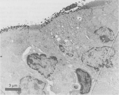
Recent evidence indicates that ileal Peyer's patches represent the main infection route for Yersinia enterocolitica. In this study transmission and scanning electron microscopy have shown that only a small fraction of bacteria present in the lumen adhere to the follicle-associated murine epithelium with no discernible preference for either M or absorptive cells. Yersiniae attached to M cells are phagocytosed and transported from the lumen into the lamina propria. No invasion of columnar absorptive cells was observed. These data, in combination with recently published reports, indicate that the involvement of M cells is a common step in bacterial invasion of Peyer's patches.

摘要

最近的证据表明,回肠派尔集合淋巴结是小肠结肠炎耶尔森菌的主要感染途径。在本研究中,透射电子显微镜和扫描电子显微镜显示,肠腔中仅一小部分细菌黏附于滤泡相关的鼠上皮细胞,对M细胞或吸收细胞均无明显偏好。黏附于M细胞的耶尔森菌被吞噬并从肠腔转运至固有层。未观察到柱状吸收细胞被侵袭。这些数据与最近发表的报告相结合,表明M细胞的参与是细菌侵袭派尔集合淋巴结的一个共同步骤。

https://cdn.ncbi.nlm.nih.gov/pmc/blobs/25d9/1378659/a5345fd66e7e/gut00603-0070-b.jpg
https://cdn.ncbi.nlm.nih.gov/pmc/blobs/25d9/1378659/e9cd1f3dc840/gut00603-0068-a.jpg
https://cdn.ncbi.nlm.nih.gov/pmc/blobs/25d9/1378659/df56cd451916/gut00603-0068-b.jpg
https://cdn.ncbi.nlm.nih.gov/pmc/blobs/25d9/1378659/1ef5bc8f81d5/gut00603-0068-c.jpg
https://cdn.ncbi.nlm.nih.gov/pmc/blobs/25d9/1378659/086ca9866ed9/gut00603-0069-a.jpg
https://cdn.ncbi.nlm.nih.gov/pmc/blobs/25d9/1378659/2cbee1706ff0/gut00603-0069-b.jpg
https://cdn.ncbi.nlm.nih.gov/pmc/blobs/25d9/1378659/2daff6ee13cf/gut00603-0070-a.jpg
https://cdn.ncbi.nlm.nih.gov/pmc/blobs/25d9/1378659/a5345fd66e7e/gut00603-0070-b.jpg

相似文献

1
Involvement of M cells in the bacterial invasion of Peyer's patches: a common mechanism shared by Yersinia enterocolitica and other enteroinvasive bacteria.M细胞参与派尔集合淋巴结的细菌侵袭:小肠结肠炎耶尔森菌和其他肠道侵袭性细菌共有的一种常见机制。
Gut. 1990 Sep;31(9):1011-5. doi: 10.1136/gut.31.9.1011.
2
Penetration of M cells and destruction of Peyer's patches by Yersinia enterocolitica: an ultrastructural and histological study.小肠结肠炎耶尔森菌对M细胞的穿透及派伊尔结的破坏:一项超微结构和组织学研究
J Med Microbiol. 1996 Apr;44(4):285-94. doi: 10.1099/00222615-44-4-285.
3
Selective association and transport of Campylobacter jejuni through M cells of rabbit Peyer's patches.空肠弯曲菌通过兔派伊尔结M细胞的选择性黏附与转运
Can J Microbiol. 1988 Oct;34(10):1142-7. doi: 10.1139/m88-201.
4
Morphology of intestinal colonization of Yersinia enterocolitica serovar O3 in mice.
J Vet Med Sci. 1999 Oct;61(10):1171-3. doi: 10.1292/jvms.61.1171.
5
The early dynamic response of the calf ileal epithelium to Salmonella typhimurium.小牛回肠上皮对鼠伤寒沙门氏菌的早期动态反应。
Vet Pathol. 1997 Sep;34(5):369-86. doi: 10.1177/030098589703400501.
6
Cytopathogenic effect of Salmonella typhi GIFU 10007 on M cells of murine ileal Peyer's patches in ligated ileal loops: an ultrastructural study.伤寒沙门菌GIFU 10007对结扎回肠袢中小鼠回肠派伊尔结M细胞的细胞病变效应:一项超微结构研究
Microbiol Immunol. 1986;30(12):1225-37. doi: 10.1111/j.1348-0421.1986.tb03055.x.
7
Yersinia enterocolitica infection of mice reveals clonal invasion and abscess formation.小鼠小肠结肠炎耶尔森菌感染显示出克隆性侵袭和脓肿形成。
Infect Immun. 2007 Aug;75(8):3802-11. doi: 10.1128/IAI.00419-07. Epub 2007 Jun 11.
8
Invasion and dissemination of Yersinia enterocolitica in the mouse infection model.小肠结肠炎耶尔森菌在小鼠感染模型中的侵袭与播散
Adv Exp Med Biol. 2007;603:279-85. doi: 10.1007/978-0-387-72124-8_25.
9
Salmonella typhimurium initiates murine infection by penetrating and destroying the specialized epithelial M cells of the Peyer's patches.鼠伤寒沙门氏菌通过穿透并破坏派尔集合淋巴结的特化上皮M细胞引发小鼠感染。
J Exp Med. 1994 Jul 1;180(1):15-23. doi: 10.1084/jem.180.1.15.
10
Toll-like receptor 2 is critical for induction of Reg3 beta expression and intestinal clearance of Yersinia pseudotuberculosis.Toll样受体2对于诱导Reg3β表达和清除肠道中的假结核耶尔森菌至关重要。
Gut. 2009 Jun;58(6):771-6. doi: 10.1136/gut.2008.168443. Epub 2009 Jan 27.

引用本文的文献

1
Functional Identification and Genetic Analysis of O-Antigen Gene Clusters of Food-Borne Pathogen O:10 and Other Uncommon Serotypes, Further Revealing Their Virulence Profiles.食源性病原体 O:10 及其它罕见血清型 O 抗原基因簇的功能鉴定与遗传分析,进一步揭示其毒力特征。
J Microbiol Biotechnol. 2024 Aug 28;34(8):1599-1608. doi: 10.4014/jmb.2402.02044. Epub 2024 Jul 15.
2
The role of goblet cells and mucus in intestinal homeostasis.杯状细胞和黏液在肠道稳态中的作用。
Nat Rev Gastroenterol Hepatol. 2022 Dec;19(12):785-803. doi: 10.1038/s41575-022-00675-x. Epub 2022 Sep 12.
3
Pathogenic Exploitation of Lymphatic Vessels.

本文引用的文献

1
Characterization of plasmids and plasmid-associated determinants of Yersinia enterocolitica pathogenesis.小肠结肠炎耶尔森菌致病性质粒及质粒相关决定因素的特征分析
Infect Immun. 1981 Feb;31(2):775-82. doi: 10.1128/iai.31.2.775-782.1981.
2
Serum resistance associated with virulence in Yersinia enterocolitica.小肠结肠炎耶尔森菌中与毒力相关的血清抗性。
Infect Immun. 1982 Feb;35(2):605-11. doi: 10.1128/iai.35.2.605-611.1982.
3
[Intestinal yersinia infection: pathogenesis, clinical course, epidemiology and diagnosis].[肠道耶尔森菌感染:发病机制、临床病程、流行病学及诊断]
淋巴管的发病机制。
Cells. 2022 Mar 12;11(6):979. doi: 10.3390/cells11060979.
4
Retinoic Acid and Lymphotoxin Signaling Promote Differentiation of Human Intestinal M Cells.维甲酸和淋巴毒素信号促进人肠道 M 细胞的分化。
Gastroenterology. 2020 Jul;159(1):214-226.e1. doi: 10.1053/j.gastro.2020.03.053. Epub 2020 Apr 1.
5
Dissemination Plays a Critical Role in Pathogenesis.传播在发病机制中起着关键作用。
Front Cell Infect Microbiol. 2020 Feb 25;10:65. doi: 10.3389/fcimb.2020.00065. eCollection 2020.
6
Iron availability and oxygen tension regulate the Yersinia Ysc type III secretion system to enable disseminated infection.铁的可用性和氧张力调节耶尔森氏菌 Ysc 型 III 型分泌系统,以实现播散性感染。
PLoS Pathog. 2019 Dec 23;15(12):e1008001. doi: 10.1371/journal.ppat.1008001. eCollection 2019 Dec.
7
The Most Important Virulence Markers of and Their Role during Infection.[病原体名称]最重要的毒力标志物及其在感染过程中的作用。 需注意,原文中“of”后面缺少具体内容,这里补充了“[病原体名称]”以使译文更完整通顺,但实际翻译时应根据准确的原文信息进行。
Genes (Basel). 2018 May 3;9(5):235. doi: 10.3390/genes9050235.
8
Allograft inflammatory factor 1 is a regulator of transcytosis in M cells.同种异体炎症因子 1 是 M 细胞易位的调节剂。
Nat Commun. 2017 Feb 22;8:14509. doi: 10.1038/ncomms14509.
9
Escherichia albertii, a novel human enteropathogen, colonizes rat enterocytes and translocates to extra-intestinal sites.阿尔伯特埃希氏菌,一种新型人类肠道病原体,可定殖于大鼠肠上皮细胞并转移至肠外部位。
PLoS One. 2017 Feb 8;12(2):e0171385. doi: 10.1371/journal.pone.0171385. eCollection 2017.
10
Manipulation of host membranes by the bacterial pathogens Listeria, Francisella, Shigella and Yersinia.细菌病原体李斯特菌、弗朗西斯菌、志贺菌和耶尔森菌对宿主细胞膜的操控。
Semin Cell Dev Biol. 2016 Dec;60:155-167. doi: 10.1016/j.semcdb.2016.07.019. Epub 2016 Jul 19.
Immun Infekt. 1982 Sep;10(5):180-6.
4
A new method using hexamethyldisilazane for preparation of soft insect tissues for scanning electron microscopy.一种使用六甲基二硅氮烷制备用于扫描电子显微镜的昆虫软组织的新方法。
Stain Technol. 1983 Nov;58(6):347-51. doi: 10.3109/10520298309066811.
5
The membranous epithelial (M) cell and the mucosal immune system.膜性上皮(M)细胞与黏膜免疫系统。
Annu Rev Med. 1984;35:95-112. doi: 10.1146/annurev.me.35.020184.000523.
6
Intestinal M cells: a pathway for entry of reovirus into the host.肠道M细胞:呼肠孤病毒进入宿主的一条途径。
Science. 1981 Apr 24;212(4493):471-2. doi: 10.1126/science.6259737.
7
Specific adherence of Escherichia coli (strain RDEC-1) to membranous (M) cells of the Peyer's patch in Escherichia coli diarrhea in the rabbit.兔大肠杆菌腹泻中大肠杆菌(RDEC-1菌株)对派伊尔结膜细胞的特异性黏附
J Clin Invest. 1983 Jan;71(1):1-8. doi: 10.1172/jci110737.
8
The pathogenesis of Yersinia enterocolitica infection in gnotobiotic piglets.悉生猪仔小肠结肠炎耶尔森菌感染的发病机制
J Med Microbiol. 1985 Jun;19(3):297-308. doi: 10.1099/00222615-19-3-297.
9
A simple adult-mouse test for tissue invasiveness in Yersinia enterocolitica strains of low experimental virulence.一种针对低实验毒力小肠结肠炎耶尔森氏菌菌株组织侵袭性的简易成年小鼠测试。
J Med Microbiol. 1985 Apr;19(2):237-46. doi: 10.1099/00222615-19-2-237.
10
Invasiveness of Yersinia enterocolitica lacking the virulence plasmid: an in-vivo study.
J Med Microbiol. 1987 Nov;24(3):219-26. doi: 10.1099/00222615-24-3-219.