• 文献检索
  • 文档翻译
  • 深度研究
  • 学术资讯
  • Suppr Zotero 插件Zotero 插件
  • 邀请有礼
  • 套餐&价格
  • 历史记录
应用&插件
Suppr Zotero 插件Zotero 插件浏览器插件Mac 客户端Windows 客户端微信小程序
定价
高级版会员购买积分包购买API积分包
服务
文献检索文档翻译深度研究API 文档MCP 服务
关于我们
关于 Suppr公司介绍联系我们用户协议隐私条款
关注我们

Suppr 超能文献

核心技术专利:CN118964589B侵权必究
粤ICP备2023148730 号-1Suppr @ 2026

文献检索

告别复杂PubMed语法,用中文像聊天一样搜索,搜遍4000万医学文献。AI智能推荐,让科研检索更轻松。

立即免费搜索

文件翻译

保留排版,准确专业,支持PDF/Word/PPT等文件格式,支持 12+语言互译。

免费翻译文档

深度研究

AI帮你快速写综述,25分钟生成高质量综述,智能提取关键信息,辅助科研写作。

立即免费体验

细胞结构介导 DivIVA 超微结构,并调节枯草芽孢杆菌中 Min 活性。

Cellular architecture mediates DivIVA ultrastructure and regulates min activity in Bacillus subtilis.

机构信息

Laboratory of Molecular Biology, National Cancer Institute, National Institutes of Health, Bethesda, Maryland, USA.

出版信息

mBio. 2011 Nov 22;2(6). doi: 10.1128/mBio.00257-11. Print 2011.

DOI:10.1128/mBio.00257-11
PMID:22108385
原文链接:https://pmc.ncbi.nlm.nih.gov/articles/PMC3225972/
Abstract

UNLABELLED

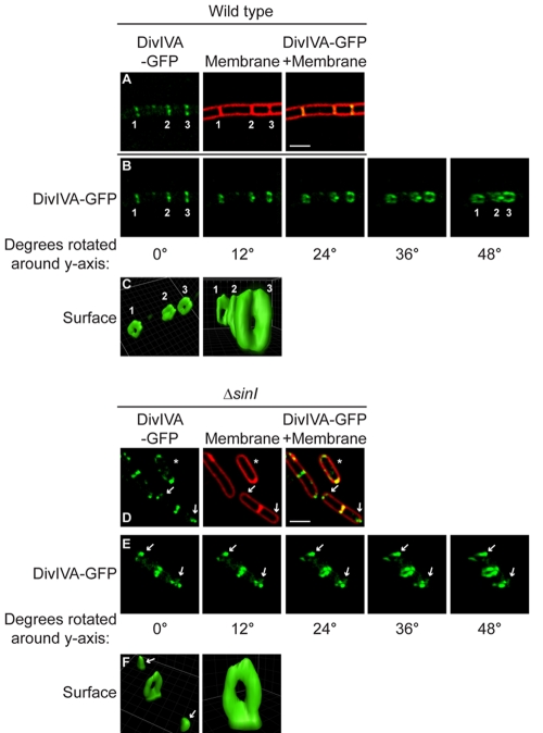
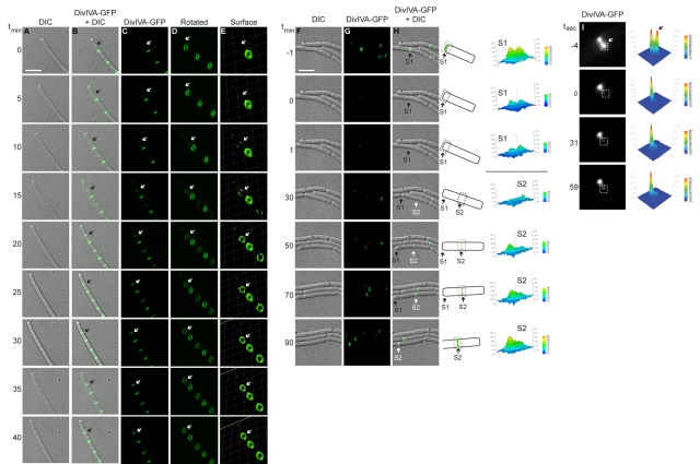
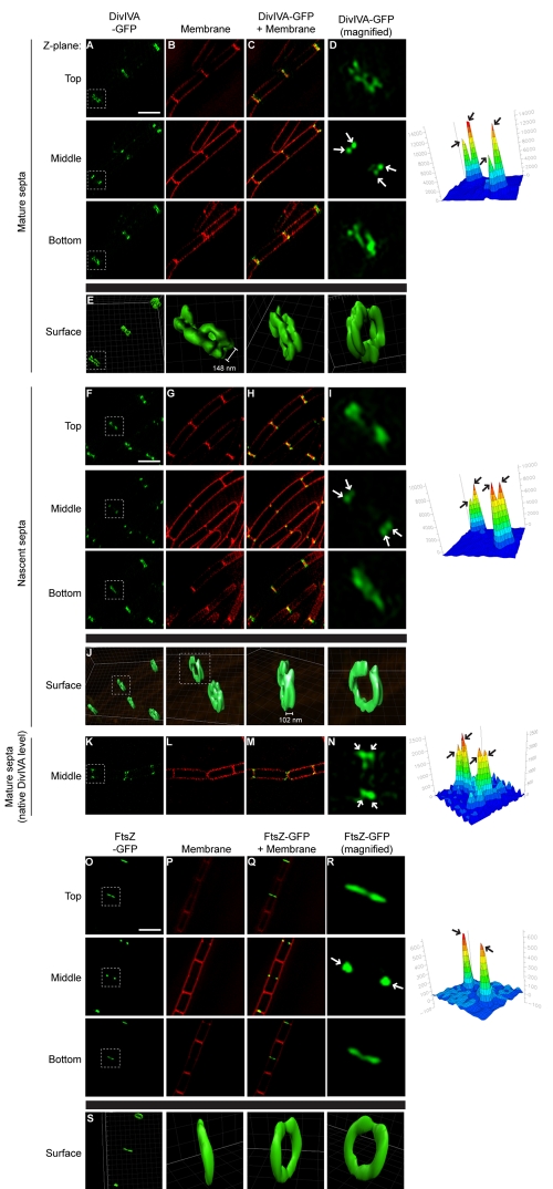
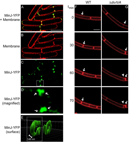
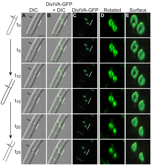
The assembly of the cell division machinery at midcell is a critical step of cytokinesis. Many rod-shaped bacteria position septa using nucleoid occlusion, which prevents division over the chromosome, and the Min system, which prevents division near the poles. Here we examined the in vivo assembly of the Bacillus subtilis MinCD targeting proteins DivIVA, a peripheral membrane protein that preferentially localizes to negatively curved membranes and resembles eukaryotic tropomyosins, and MinJ, which recruits MinCD to DivIVA. We used structured illumination microscopy to demonstrate that both DivIVA and MinJ localize as double rings that flank the septum and first appear early in septal biosynthesis. The subsequent recruitment of MinCD to these double rings would separate the Min proteins from their target, FtsZ, spatially regulating Min activity and allowing continued cell division. Curvature-based localization would also provide temporal regulation, since DivIVA and the Min proteins would localize to midcell after the onset of division. We use time-lapse microscopy and fluorescence recovery after photobleaching to demonstrate that DivIVA rings are highly stable and are constructed from newly synthesized DivIVA molecules. After cell division, DivIVA rings appear to collapse into patches at the rounded cell poles of separated cells, with little or no incorporation of newly synthesized subunits. Thus, changes in cell architecture mediate both the initial recruitment of DivIVA to sites of cell division and the subsequent collapse of these rings into patches (or rings of smaller diameter), while curvature-based localization of DivIVA spatially and temporally regulates Min activity.

IMPORTANCE

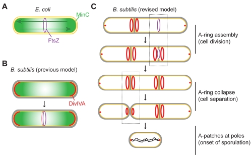
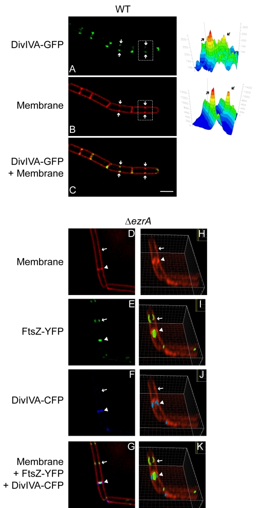
The Min systems of Escherichia coli and Bacillus subtilis both inhibit FtsZ assembly, but one key difference between these two species is that whereas the E. coli Min proteins localize to the poles, the B. subtilis proteins localize to nascent division sites by interaction with DivIVA and MinJ. It is unclear how MinC activity at midcell is regulated to prevent it from interfering with FtsZ engaged in medial cell division. We used superresolution microscopy to demonstrate that DivIVA and MinJ, which localize MinCD, assemble double rings that flank active division sites and septa. This curvature-based localization mechanism holds MinCD away from the FtsZ ring at midcell, and we propose that this spatial organization is the primary mechanism by which MinC activity is regulated to allow division at midcell. Curvature-based localization also conveys temporal regulation, since it ensures that MinC localizes after the onset of division.

摘要

未加标签

细胞分裂装置在细胞中部的组装是细胞分裂的关键步骤。许多杆状细菌使用类核阻塞来定位隔膜,类核阻塞阻止了染色体的分裂,而 Min 系统则防止了靠近极的分裂。在这里,我们研究了枯草芽孢杆菌 MinCD 靶向蛋白 DivIVA 的体内组装,DivIVA 是一种优先定位于负曲率膜的外周膜蛋白,类似于真核细胞的原肌球蛋白,以及 MinJ,它将 MinCD 招募到 DivIVA。我们使用结构光照明显微镜证明 DivIVA 和 MinJ 都定位于隔膜两侧的双环中,并且这些双环最早出现在隔膜生物合成的早期。随后 MinCD 对这些双环的募集将 Min 蛋白与其靶标 FtsZ 分离,从而在空间上调节 Min 活性并允许细胞继续分裂。基于曲率的定位还提供了时间调节,因为 DivIVA 和 Min 蛋白在细胞分裂开始后才会定位于细胞中部。我们使用延时显微镜和光漂白后荧光恢复来证明 DivIVA 环非常稳定,并且由新合成的 DivIVA 分子构建。细胞分裂后,DivIVA 环似乎在分离细胞的圆形细胞极处崩溃成斑块,很少或没有新合成的亚基掺入。因此,细胞结构的变化介导了 DivIVA 最初被招募到细胞分裂部位,以及随后这些环崩溃成斑块(或直径较小的环),而 DivIVA 的基于曲率的定位在空间和时间上调节 Min 活性。

重要性

大肠杆菌和枯草芽孢杆菌的 Min 系统都抑制 FtsZ 组装,但这两个物种之间的一个关键区别是,大肠杆菌的 Min 蛋白定位于极,而枯草芽孢杆菌的蛋白通过与 DivIVA 和 MinJ 相互作用定位于新的分裂部位。MinC 在细胞中部的活性如何被调节以防止其干扰参与中部分裂的 FtsZ 尚不清楚。我们使用超分辨率显微镜证明,定位 MinCD 的 DivIVA 和 MinJ 组装了定位于活跃分裂部位和隔膜两侧的双环。这种基于曲率的定位机制将 MinCD 与 FtsZ 环在细胞中部隔开,我们提出这种空间组织是调节 MinC 活性以允许在细胞中部进行分裂的主要机制。基于曲率的定位也传递了时间调节,因为它确保 MinC 在细胞分裂开始后定位。

https://cdn.ncbi.nlm.nih.gov/pmc/blobs/ed6b/3225972/2f41dcf1fa9d/mbo0061111980007.jpg
https://cdn.ncbi.nlm.nih.gov/pmc/blobs/ed6b/3225972/2fbfdc75dd51/mbo0061111980001.jpg
https://cdn.ncbi.nlm.nih.gov/pmc/blobs/ed6b/3225972/5a4b0f09e63e/mbo0061111980002.jpg
https://cdn.ncbi.nlm.nih.gov/pmc/blobs/ed6b/3225972/7bb60184356b/mbo0061111980003.jpg
https://cdn.ncbi.nlm.nih.gov/pmc/blobs/ed6b/3225972/6be982680211/mbo0061111980004.jpg
https://cdn.ncbi.nlm.nih.gov/pmc/blobs/ed6b/3225972/64fd608c8b38/mbo0061111980005.jpg
https://cdn.ncbi.nlm.nih.gov/pmc/blobs/ed6b/3225972/835db1473951/mbo0061111980006.jpg
https://cdn.ncbi.nlm.nih.gov/pmc/blobs/ed6b/3225972/2f41dcf1fa9d/mbo0061111980007.jpg

相似文献

1
Cellular architecture mediates DivIVA ultrastructure and regulates min activity in Bacillus subtilis.细胞结构介导 DivIVA 超微结构,并调节枯草芽孢杆菌中 Min 活性。
mBio. 2011 Nov 22;2(6). doi: 10.1128/mBio.00257-11. Print 2011.
2
Bacillus subtilis MinC destabilizes FtsZ-rings at new cell poles and contributes to the timing of cell division.枯草芽孢杆菌MinC会破坏新细胞极处的FtsZ环,并对细胞分裂的时间安排有影响。
Genes Dev. 2008 Dec 15;22(24):3475-88. doi: 10.1101/gad.1732408.
3
Septal membrane localization by C-terminal amphipathic α-helices of MinD in Bacillus subtilis mutant cells lacking MinJ or DivIVA.在缺乏MinJ或DivIVA的枯草芽孢杆菌突变细胞中,MinD的C端两亲性α螺旋对隔膜的定位作用
Genes Genet Syst. 2017 Oct 18;92(2):81-98. doi: 10.1266/ggs.16-00054. Epub 2017 Jun 30.
4
A novel component of the division-site selection system of Bacillus subtilis and a new mode of action for the division inhibitor MinCD.枯草芽孢杆菌分裂位点选择系统的一种新组分以及分裂抑制剂MinCD的一种新作用模式。
Mol Microbiol. 2008 Dec;70(6):1556-69. doi: 10.1111/j.1365-2958.2008.06501.x. Epub 2008 Oct 23.
5
Selection of the midcell division site in Bacillus subtilis through MinD-dependent polar localization and activation of MinC.通过MinD依赖的极性定位和MinC激活来选择枯草芽孢杆菌中的细胞中部分裂位点。
Mol Microbiol. 1999 Jul;33(1):84-96. doi: 10.1046/j.1365-2958.1999.01450.x.
6
MinJ (YvjD) is a topological determinant of cell division in Bacillus subtilis.MinJ(YvjD)是枯草芽孢杆菌细胞分裂的拓扑学决定因素。
Mol Microbiol. 2008 Dec;70(5):1166-79. doi: 10.1111/j.1365-2958.2008.06469.x. Epub 2008 Oct 2.
7
Protein-protein interaction domains of Bacillus subtilis DivIVA.枯草芽孢杆菌 DivIVA 的蛋白质-蛋白质相互作用结构域。
J Bacteriol. 2013 Mar;195(5):1012-21. doi: 10.1128/JB.02171-12. Epub 2012 Dec 21.
8
Imaging DivIVA dynamics using photo-convertible and activatable fluorophores in Bacillus subtilis.利用枯草芽孢杆菌中光转化和可激活荧光染料来成像 DivIVA 动力学。
Front Microbiol. 2014 Feb 18;5:59. doi: 10.3389/fmicb.2014.00059. eCollection 2014.
9
How Do MinC-D Copolymers Act on Z-Ring Localization Regulation? A New Model of Min System.MinC-D共聚物如何作用于Z环定位调控?Min系统的新模型。
Front Microbiol. 2022 Apr 15;13:841171. doi: 10.3389/fmicb.2022.841171. eCollection 2022.
10
Early targeting of Min proteins to the cell poles in germinated spores of Bacillus subtilis: evidence for division apparatus-independent recruitment of Min proteins to the division site.枯草芽孢杆菌萌发孢子中Min蛋白早期定位于细胞两极:Min蛋白在不依赖分裂装置的情况下募集到分裂位点的证据。
Mol Microbiol. 2003 Jan;47(1):37-48. doi: 10.1046/j.1365-2958.2003.03253.x.

引用本文的文献

1
Phenotypic plasticity in cell elongation among closely related bacterial species.亲缘关系相近的细菌物种间细胞伸长的表型可塑性。
Nat Commun. 2025 Jun 2;16(1):5099. doi: 10.1038/s41467-025-60005-y.
2
How do spherical bacteria regulate cell division?球形细菌如何调节细胞分裂?
Biochem Soc Trans. 2025 Apr 17;53(2):447-60. doi: 10.1042/BST20240956.
3
Exploration and analytical techniques for membrane curvature-sensing proteins in bacteria.细菌中膜曲率感知蛋白的探索与分析技术

本文引用的文献

1
The Min oscillator uses MinD-dependent conformational changes in MinE to spatially regulate cytokinesis.Min 振荡器利用 MinD 依赖性的 MinE 构象变化来空间调节细胞分裂。
Cell. 2011 Aug 5;146(3):396-407. doi: 10.1016/j.cell.2011.06.042.
2
FtsZ in bacterial cytokinesis: cytoskeleton and force generator all in one.细菌胞质分裂中的 FtsZ:细胞骨架和力发生器合二为一。
Microbiol Mol Biol Rev. 2010 Dec;74(4):504-28. doi: 10.1128/MMBR.00021-10.
3
Advances in understanding E. coli cell fission.对大肠杆菌细胞分裂的认识进展。
J Bacteriol. 2025 Apr 17;207(4):e0048224. doi: 10.1128/jb.00482-24. Epub 2025 Mar 26.
4
GpsB interacts with FtsZ in multiple species and may serve as an accessory Z-ring anchor.GpsB在多个物种中与FtsZ相互作用,可能作为一种辅助性Z环锚定蛋白。
Mol Biol Cell. 2025 Jan 1;36(1):ar10. doi: 10.1091/mbc.E24-07-0302. Epub 2024 Nov 27.
5
A dispensable SepIVA orthologue in Streptomyces venezuelae is associated with polar growth and not cell division.委内瑞拉链霉菌中一个可有可无的 SepIVA 同源物与极性生长而非细胞分裂有关。
BMC Microbiol. 2024 Nov 18;24(1):481. doi: 10.1186/s12866-024-03625-6.
6
PcdA promotes orthogonal division plane selection in Staphylococcus aureus.PcdA 促进金黄色葡萄球菌的正交分裂平面选择。
Nat Microbiol. 2024 Nov;9(11):2997-3012. doi: 10.1038/s41564-024-01821-8. Epub 2024 Oct 28.
7
Polymer dynamics of Alp7A reveals how two critical concentrations govern assembly of dynamically unstable actin-like proteins.Alp7A 的聚合物动力学揭示了两种关键浓度如何控制动态不稳定肌动蛋白样蛋白的组装。
Mol Biol Cell. 2024 Nov 1;35(11):ar145. doi: 10.1091/mbc.E23-11-0440. Epub 2024 Sep 25.
8
Cell division machinery drives cell-specific gene activation during differentiation in .细胞分裂机制在 分化过程中驱动细胞特异性基因激活。
Proc Natl Acad Sci U S A. 2024 Mar 26;121(13):e2400584121. doi: 10.1073/pnas.2400584121. Epub 2024 Mar 19.
9
Distinct dynamics and proximity networks of hub proteins at the prey-invading cell pole in a predatory bacterium.在一种捕食性细菌中,位于猎物入侵细胞极的枢纽蛋白的不同动力学和临近网络。
J Bacteriol. 2024 Apr 18;206(4):e0001424. doi: 10.1128/jb.00014-24. Epub 2024 Mar 12.
10
Comparing divisome organization between vegetative and sporulating at the nanoscale using DNA-PAINT.利用 DNA-PAINT 在纳米尺度上比较营养体和孢子形成期的分隔体组织。
Sci Adv. 2024 Jan 12;10(2):eadk5847. doi: 10.1126/sciadv.adk5847. Epub 2024 Jan 10.
Curr Opin Microbiol. 2010 Dec;13(6):730-7. doi: 10.1016/j.mib.2010.09.015. Epub 2010 Oct 11.
4
Changes in the Min oscillation pattern before and after cell birth.细胞分裂前后 Min 振荡模式的变化。
J Bacteriol. 2010 Aug;192(16):4134-42. doi: 10.1128/JB.00364-10. Epub 2010 Jun 11.
5
Features critical for membrane binding revealed by DivIVA crystal structure.DivIVA 晶体结构揭示了对膜结合至关重要的特征。
EMBO J. 2010 Jun 16;29(12):1988-2001. doi: 10.1038/emboj.2010.99. Epub 2010 May 25.
6
Protein subcellular localization in bacteria.细菌中的蛋白质亚细胞定位。
Cold Spring Harb Perspect Biol. 2010 Apr;2(4):a000307. doi: 10.1101/cshperspect.a000307. Epub 2010 Mar 3.
7
The MinCDJ system in Bacillus subtilis prevents minicell formation by promoting divisome disassembly.枯草芽孢杆菌中的 MinCDJ 系统通过促进分裂体的解体来防止小型细胞的形成。
PLoS One. 2010 Mar 24;5(3):e9850. doi: 10.1371/journal.pone.0009850.
8
Why and how bacteria localize proteins.细菌为何以及如何定位蛋白质。
Science. 2009 Nov 27;326(5957):1225-8. doi: 10.1126/science.1175685.
9
Sculpting the bacterial cell.塑造细菌细胞。
Curr Biol. 2009 Sep 15;19(17):R812-22. doi: 10.1016/j.cub.2009.06.033.
10
Division site selection in rod-shaped bacteria.杆状菌的分裂位点选择。
Curr Opin Microbiol. 2009 Dec;12(6):683-8. doi: 10.1016/j.mib.2009.10.002. Epub 2009 Nov 1.