Barreto-Bergter Eliana, Sassaki Guilherme L, de Souza Lauro M
Instituto de Microbiologia Paulo de Góes, Universidade Federal do Rio de Janeiro Rio de Janeiro, Brazil.
Front Microbiol. 2011 Dec 5;2:239. doi: 10.3389/fmicb.2011.00239. eCollection 2011.
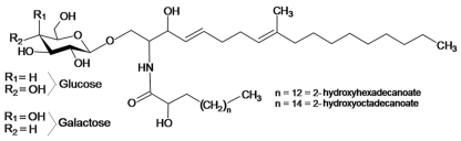
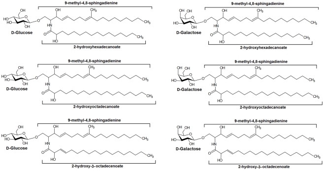
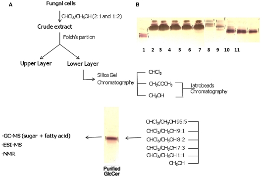
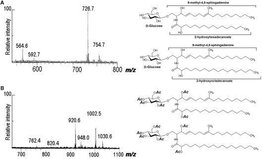
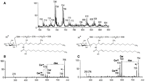
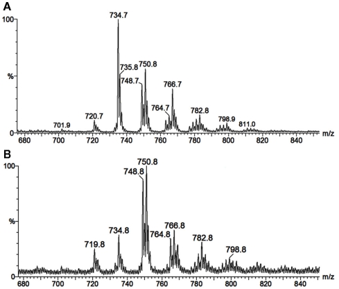
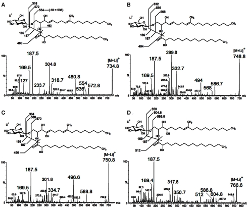
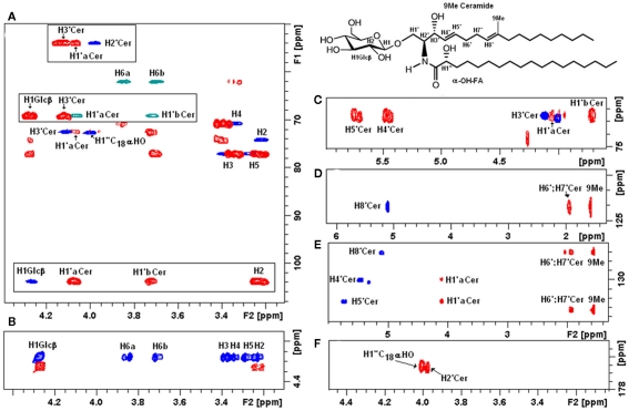
Of the ceramide monohexosides (CMHs), gluco- and galactosyl-ceramides are the main neutral glycosphingolipids expressed in fungal cells. Their structural determination is greatly dependent on the use of mass spectrometric techniques, including fast atom bombardment-mass spectrometry, electrospray ionization, and energy collision-induced dissociation mass spectrometry. Nuclear magnetic resonance has also been used successfully. Such a combination of techniques, combined with classical analytical separation, such as high-performance thin layer chromatography and column chromatography, has led to the structural elucidation of a great number of fungal CMHs. The structure of fungal CMH is conserved among fungal species and consists of a glucose or galactose residue attached to a ceramide moiety containing 9-methyl-4,8-sphingadienine with an amidic linkage to hydroxylated fatty acids, most commonly having 16 or 18 carbon atoms and unsaturation between C-3 and C-4. Along with their unique structural characteristics, fungal CMHs have a peculiar subcellular distribution and striking biological properties. Fungal cerebrosides were also characterized as antigenic molecules directly or indirectly involved in cell growth or differentiation in Schizophyllum commune, Cryptococcus neoformans, Pseudallescheria boydii, Candida albicans, Aspergillus nidulans, Aspergillus fumigatus, and Colletotrichum gloeosporioides. Besides classical techniques for cerebroside (CMH) analysis, we now describe new approaches, combining conventional thin layer chromatography and mass spectrometry, as well as emerging technologies for subcellular localization and distribution of glycosphingolipids by secondary ion mass spectrometry and imaging matrix-assisted laser desorption ionization time-of-flight.
在神经酰胺单己糖苷(CMHs)中,葡萄糖基神经酰胺和半乳糖基神经酰胺是真菌细胞中表达的主要中性糖鞘脂。它们的结构测定在很大程度上依赖于质谱技术的应用,包括快原子轰击质谱、电喷雾电离和能量碰撞诱导解离质谱。核磁共振也已成功应用。这种技术组合,再加上经典的分析分离方法,如高效薄层色谱和柱色谱,已使大量真菌CMHs的结构得以阐明。真菌CMH的结构在真菌物种中是保守的,由一个葡萄糖或半乳糖残基连接到一个神经酰胺部分组成,该神经酰胺部分含有9-甲基-4,8-鞘氨二烯,与羟基化脂肪酸通过酰胺键相连,这些脂肪酸最常见的是含有16或18个碳原子,且在C-3和C-4之间存在不饱和键。除了其独特的结构特征外,真菌CMHs还具有特殊的亚细胞分布和显著的生物学特性。在裂褶菌、新型隐球菌、博伊德假阿利什菌、白色念珠菌、烟曲霉、黄曲霉和炭疽菌中,真菌脑苷脂也被表征为直接或间接参与细胞生长或分化的抗原分子。除了用于脑苷脂(CMH)分析的经典技术外,我们现在还描述了结合传统薄层色谱和质谱的新方法,以及通过二次离子质谱和成像基质辅助激光解吸电离飞行时间质谱对糖鞘脂进行亚细胞定位和分布的新兴技术。