• 文献检索
  • 文档翻译
  • 深度研究
  • 学术资讯
  • Suppr Zotero 插件Zotero 插件
  • 邀请有礼
  • 套餐&价格
  • 历史记录
应用&插件
Suppr Zotero 插件Zotero 插件浏览器插件Mac 客户端Windows 客户端微信小程序
定价
高级版会员购买积分包购买API积分包
服务
文献检索文档翻译深度研究API 文档MCP 服务
关于我们
关于 Suppr公司介绍联系我们用户协议隐私条款
关注我们

Suppr 超能文献

核心技术专利:CN118964589B侵权必究
粤ICP备2023148730 号-1Suppr @ 2026

文献检索

告别复杂PubMed语法,用中文像聊天一样搜索,搜遍4000万医学文献。AI智能推荐,让科研检索更轻松。

立即免费搜索

文件翻译

保留排版,准确专业,支持PDF/Word/PPT等文件格式,支持 12+语言互译。

免费翻译文档

深度研究

AI帮你快速写综述,25分钟生成高质量综述,智能提取关键信息,辅助科研写作。

立即免费体验

验证一种动脉迂曲度测量方法及其在高血压临床患者队列中的应用。

Validation of an arterial tortuosity measure with application to hypertension collection of clinical hypertensive patients.

机构信息

Utah Center for Advanced Imaging Research, Department of Radiology, University of Utah, 729 Arapeen Drive, Salt Lake City, UT 84108, USA.

出版信息

BMC Bioinformatics. 2011 Oct 18;12 Suppl 10(Suppl 10):S15. doi: 10.1186/1471-2105-12-S10-S15.

DOI:10.1186/1471-2105-12-S10-S15
PMID:22166145
原文链接:https://pmc.ncbi.nlm.nih.gov/articles/PMC3236837/
Abstract

BACKGROUND

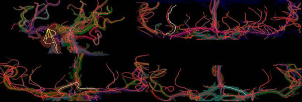
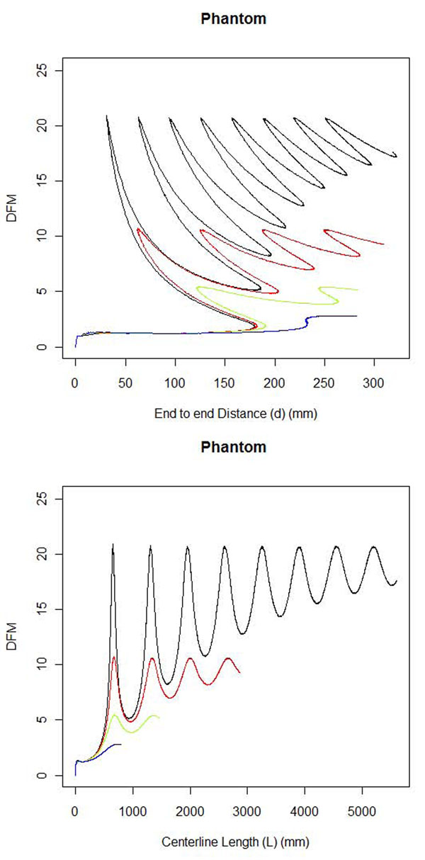
Hypertension may increase tortuosity or twistedness of arteries. We applied a centerline extraction algorithm and tortuosity metric to magnetic resonance angiography (MRA) brain images to quantitatively measure the tortuosity of arterial vessel centerlines. The most commonly used arterial tortuosity measure is the distance factor metric (DFM). This study tested a DFM based measurement's ability to detect increases in arterial tortuosity of hypertensives using existing images. Existing images presented challenges such as different resolutions which may affect the tortuosity measurement, different depths of the area imaged, and different artifacts of imaging that require filtering.

METHODS

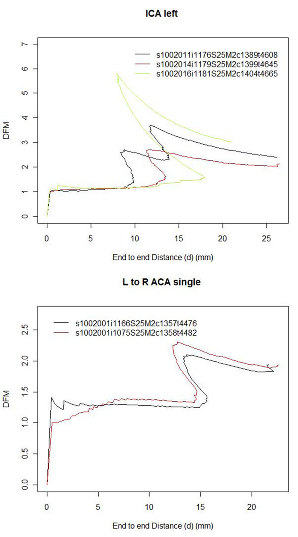
The stability and accuracy of alternative centerline algorithms was validated in numerically generated models and test brain MRA data. Existing images were gathered from previous studies and clinical medical systems by manually reading electronic medical records to identify hypertensives and negatives. Images of different resolutions were interpolated to similar resolutions. Arterial tortuosity in MRA images was measured from a DFM curve and tested on numerically generated models as well as MRA images from two hypertensive and three negative control populations. Comparisons were made between different resolutions, different filters, hypertensives versus negatives, and different negative controls.

RESULTS

In tests using numerical models of a simple helix, the measured tortuosity increased as expected with more tightly coiled helices. Interpolation reduced resolution-dependent differences in measured tortuosity. The Korean hypertensive population had significantly higher arterial tortuosity than its corresponding negative control population across multiple arteries. In addition one negative control population of different ethnicity had significantly less arterial tortuosity than the other two.

CONCLUSIONS

Tortuosity can be compared between images of different resolutions by interpolating from lower to higher resolutions. Use of a universal negative control was not possible in this study. The method described here detected elevated arterial tortuosity in a hypertensive population compared to the negative control population and can be used to study this relation in other populations.

摘要

背景

高血压可能会增加动脉的迂曲或扭曲程度。我们应用中心线提取算法和迂曲度度量标准对磁共振血管造影(MRA)脑图像进行分析,以定量测量动脉中心线的迂曲程度。最常用的动脉迂曲度度量标准是距离因子度量(DFM)。本研究使用现有的图像来测试一种基于 DFM 的测量方法,以检测高血压患者动脉迂曲度的增加。现有的图像存在一些挑战,例如分辨率不同可能会影响迂曲度的测量,成像区域的深度不同,以及需要过滤的成像伪影不同。

方法

在数值生成的模型和测试脑 MRA 数据中验证了替代中心线算法的稳定性和准确性。通过手动阅读电子病历,从先前的研究和临床医疗系统中收集现有的图像,以识别高血压患者和非高血压患者。对不同分辨率的图像进行插值,以达到相似的分辨率。使用 DFM 曲线测量 MRA 图像中的动脉迂曲度,并在数值生成的模型以及来自两个高血压和三个阴性对照组的 MRA 图像上进行测试。比较了不同分辨率、不同滤波器、高血压患者与非高血压患者以及不同阴性对照组之间的差异。

结果

在使用简单螺旋的数值模型进行的测试中,随着螺旋的紧密程度增加,测量的迂曲度按预期增加。插值减少了分辨率依赖性测量迂曲度的差异。与相应的阴性对照组相比,韩国高血压患者的多个动脉迂曲度明显更高。此外,与其他两个对照组相比,一个不同种族的阴性对照组的动脉迂曲度明显较低。

结论

通过从低分辨率插值到高分辨率,可以比较不同分辨率的图像之间的迂曲度。在本研究中,使用通用的阴性对照组是不可能的。这里描述的方法检测到高血压患者与阴性对照组相比,动脉迂曲度升高,并可用于研究其他人群中的这种关系。

https://cdn.ncbi.nlm.nih.gov/pmc/blobs/a4d4/3236837/a1a3b8916ade/1471-2105-12-S10-S15-7.jpg
https://cdn.ncbi.nlm.nih.gov/pmc/blobs/a4d4/3236837/86c8acfd5735/1471-2105-12-S10-S15-4.jpg
https://cdn.ncbi.nlm.nih.gov/pmc/blobs/a4d4/3236837/cdb908bf32b1/1471-2105-12-S10-S15-5.jpg
https://cdn.ncbi.nlm.nih.gov/pmc/blobs/a4d4/3236837/a1a3b8916ade/1471-2105-12-S10-S15-7.jpg
https://cdn.ncbi.nlm.nih.gov/pmc/blobs/a4d4/3236837/86c8acfd5735/1471-2105-12-S10-S15-4.jpg
https://cdn.ncbi.nlm.nih.gov/pmc/blobs/a4d4/3236837/cdb908bf32b1/1471-2105-12-S10-S15-5.jpg
https://cdn.ncbi.nlm.nih.gov/pmc/blobs/a4d4/3236837/a1a3b8916ade/1471-2105-12-S10-S15-7.jpg

相似文献

1
Validation of an arterial tortuosity measure with application to hypertension collection of clinical hypertensive patients.验证一种动脉迂曲度测量方法及其在高血压临床患者队列中的应用。
BMC Bioinformatics. 2011 Oct 18;12 Suppl 10(Suppl 10):S15. doi: 10.1186/1471-2105-12-S10-S15.
2
Comparing performance of centerline algorithms for quantitative assessment of brain vascular anatomy.比较中心线算法在定量评估脑血管解剖结构中的性能。
Anat Rec (Hoboken). 2012 Dec;295(12):2179-90. doi: 10.1002/ar.22603. Epub 2012 Oct 12.
3
Intracranial Arterial Tortuosity in Marfan Syndrome and Loeys-Dietz Syndrome: Tortuosity Index Evaluation Is Useful in the Differential Diagnosis.马凡综合征和洛伊茨-迪茨综合征的颅内动脉迂曲:迂曲指数评估有助于鉴别诊断。
AJNR Am J Neuroradiol. 2020 Oct;41(10):1916-1922. doi: 10.3174/ajnr.A6732. Epub 2020 Aug 20.
4
Measuring tortuosity of the intracerebral vasculature from MRA images.从磁共振血管造影(MRA)图像测量脑内血管的迂曲度。
IEEE Trans Med Imaging. 2003 Sep;22(9):1163-71. doi: 10.1109/TMI.2003.816964.
5
A Novel Framework for Early Detection of Hypertension using Magnetic Resonance Angiography.基于磁共振血管成像的高血压早期检测新框架
Sci Rep. 2019 Jul 31;9(1):11105. doi: 10.1038/s41598-019-47368-1.
6
Threshold segmentation algorithm for automatic extraction of cerebral vessels from brain magnetic resonance angiography images.用于从脑磁共振血管造影图像中自动提取脑血管的阈值分割算法。
J Neurosci Methods. 2015 Feb 15;241:30-6. doi: 10.1016/j.jneumeth.2014.12.003. Epub 2014 Dec 11.
7
Automatic detection of three-dimensional vascular tree centerlines and bifurcations in high-resolution magnetic resonance angiography.在高分辨率磁共振血管造影中自动检测三维血管树中心线和分支点。
Invest Radiol. 2005 Oct;40(10):661-71. doi: 10.1097/01.rli.0000178433.32526.e0.
8
Time-of-Arrival Parametric Maps and Virtual Bolus Images Derived From Contrast-Enhanced Time-Resolved Radial Magnetic Resonance Angiography Improve the Display of Brain Arteriovenous Malformation Vascular Anatomy.基于对比增强时间分辨径向磁共振血管造影术的到达时间参数图和虚拟团注图像改善了脑动静脉畸形血管解剖结构的显示。
Invest Radiol. 2016 Nov;51(11):706-713. doi: 10.1097/RLI.0000000000000288.
9
[Magnetic resonance angiography of the vessels of the neck: the optimization of a dynamic technic during the rapid infusion of a paramagnetic contrast medium].[颈部血管的磁共振血管造影:在快速注入顺磁性造影剂过程中动态技术的优化]
Radiol Med. 1997 Oct;94(4):325-8.
10
Tortuosity in non-atherosclerotic vascular diseases is associated with age, arterial aneurysms, and hypertension.在非动脉粥样硬化性血管疾病中,迂曲与年龄、动脉瘤和高血压有关。
Orphanet J Rare Dis. 2024 Jun 7;19(1):227. doi: 10.1186/s13023-024-03231-9.

引用本文的文献

1
Tortuosity in non-atherosclerotic vascular diseases is associated with age, arterial aneurysms, and hypertension.在非动脉粥样硬化性血管疾病中,迂曲与年龄、动脉瘤和高血压有关。
Orphanet J Rare Dis. 2024 Jun 7;19(1):227. doi: 10.1186/s13023-024-03231-9.
2
Modification of cerebrovascular morphologies during different stages of life.脑血管形态在生命不同阶段的变化。
J Cereb Blood Flow Metab. 2022 Nov;42(11):2151-2160. doi: 10.1177/0271678X221111609. Epub 2022 Jul 1.
3
Evaluation of Brain Tortuosity Measurement for the Automatic Multimodal Classification of Subjects with Alzheimer's Disease.

本文引用的文献

1
Geometric variability of the abdominal aorta and its major peripheral branches.腹主动脉及其主要外周分支的几何变异。
Ann Biomed Eng. 2010 Mar;38(3):824-40. doi: 10.1007/s10439-010-9925-5. Epub 2010 Jan 20.
2
Hypertension correlates with lenticulostriate arteries visualized by 7T magnetic resonance angiography.高血压与通过7T磁共振血管造影术显示的豆纹动脉相关。
Hypertension. 2009 Nov;54(5):1050-6. doi: 10.1161/HYPERTENSIONAHA.109.140350. Epub 2009 Oct 5.
3
Imaging and analysis of lenticulostriate arteries using 7.0-Tesla magnetic resonance angiography.
评估脑扭曲度测量在阿尔茨海默病患者自动多模态分类中的应用。
Comput Intell Neurosci. 2020 Jan 29;2020:4041832. doi: 10.1155/2020/4041832. eCollection 2020.
4
Arterial Tortuosity: An Imaging Biomarker of Childhood Stroke Pathogenesis?动脉迂曲:儿童卒中发病机制的一种影像学生物标志物?
Stroke. 2016 May;47(5):1265-70. doi: 10.1161/STROKEAHA.115.011331. Epub 2016 Mar 22.
5
Quantification of tortuosity and fractal dimension of the lung vessels in pulmonary hypertension patients.量化肺动脉高压患者肺血管的迂曲度和分形维数。
PLoS One. 2014 Jan 31;9(1):e87515. doi: 10.1371/journal.pone.0087515. eCollection 2014.
6
Digital reconstruction and morphometric analysis of human brain arterial vasculature from magnetic resonance angiography.基于磁共振血管成像的人脑动脉血管的数字重建与形态计量分析。
Neuroimage. 2013 Nov 15;82:170-81. doi: 10.1016/j.neuroimage.2013.05.089. Epub 2013 May 28.
7
Proceedings of the 2012 MidSouth Computational Biology and Bioinformatics Society (MCBIOS) conference. Introduction.2012年中南计算生物学与生物信息学学会(MCBIOS)会议论文集。引言。
BMC Bioinformatics. 2012;13 Suppl 15(Suppl 15):S1. doi: 10.1186/1471-2105-13-S15-S1. Epub 2012 Sep 11.
8
Proceedings of the 2011 MidSouth Computational Biology and Bioinformatics Society (MCBIOS) conference. Introduction.2011年中南计算生物学与生物信息学学会(MCBIOS)会议论文集。引言。
BMC Bioinformatics. 2011 Oct 18;12 Suppl 10(Suppl 10):S1. doi: 10.1186/1471-2105-12-S10-S1.
使用7.0特斯拉磁共振血管造影对豆纹动脉进行成像与分析。
Magn Reson Med. 2009 Jan;61(1):136-44. doi: 10.1002/mrm.21786.
4
The effects of healthy aging on intracerebral blood vessels visualized by magnetic resonance angiography.磁共振血管造影显示健康老龄化对颅内血管的影响。
Neurobiol Aging. 2010 Feb;31(2):290-300. doi: 10.1016/j.neurobiolaging.2008.03.022.
5
Observation of the lenticulostriate arteries in the human brain in vivo using 7.0T MR angiography.使用7.0T磁共振血管造影术对人脑豆纹动脉进行活体观察。
Stroke. 2008 May;39(5):1604-6. doi: 10.1161/STROKEAHA.107.508002. Epub 2008 Mar 13.
6
Loeys-Dietz syndrome: MDCT angiography findings.洛伊斯-迪茨综合征:多层螺旋CT血管造影表现
AJR Am J Roentgenol. 2007 Jul;189(1):W29-35. doi: 10.2214/AJR.06.1316.
7
A review of micro- and macrovascular analyses in the assessment of tumor-associated vasculature as visualized by MR.磁共振成像所见肿瘤相关脉管系统评估中微血管与大血管分析的综述
Neuroimage. 2007;37 Suppl 1(Suppl 1):S116-9. doi: 10.1016/j.neuroimage.2007.03.067. Epub 2007 Apr 25.
8
Methods for three-dimensional geometric characterization of the arterial vasculature.动脉血管系统的三维几何特征描述方法。
Ann Biomed Eng. 2007 Aug;35(8):1368-81. doi: 10.1007/s10439-007-9307-9. Epub 2007 Apr 13.
9
Automatic detection of three-dimensional vascular tree centerlines and bifurcations in high-resolution magnetic resonance angiography.在高分辨率磁共振血管造影中自动检测三维血管树中心线和分支点。
Invest Radiol. 2005 Oct;40(10):661-71. doi: 10.1097/01.rli.0000178433.32526.e0.
10
A syndrome of altered cardiovascular, craniofacial, neurocognitive and skeletal development caused by mutations in TGFBR1 or TGFBR2.由TGFBR1或TGFBR2突变引起的心血管、颅面、神经认知和骨骼发育改变综合征。
Nat Genet. 2005 Mar;37(3):275-81. doi: 10.1038/ng1511. Epub 2005 Jan 30.