Wellcome Trust Centre for Neuroimaging, UCL Institute of Neurology, London, UK.
Neuroimage. 2012 Mar;60(1):83-94. doi: 10.1016/j.neuroimage.2011.11.082. Epub 2011 Dec 8.
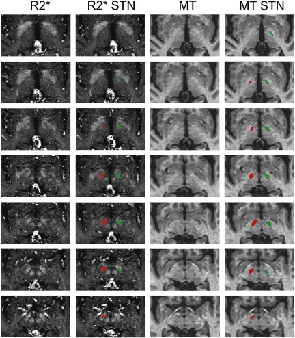
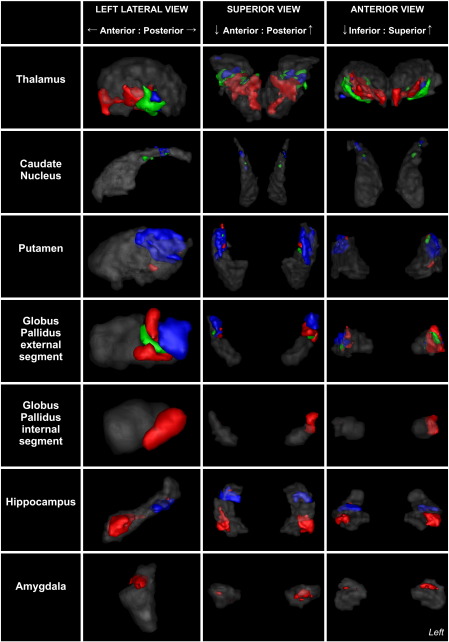
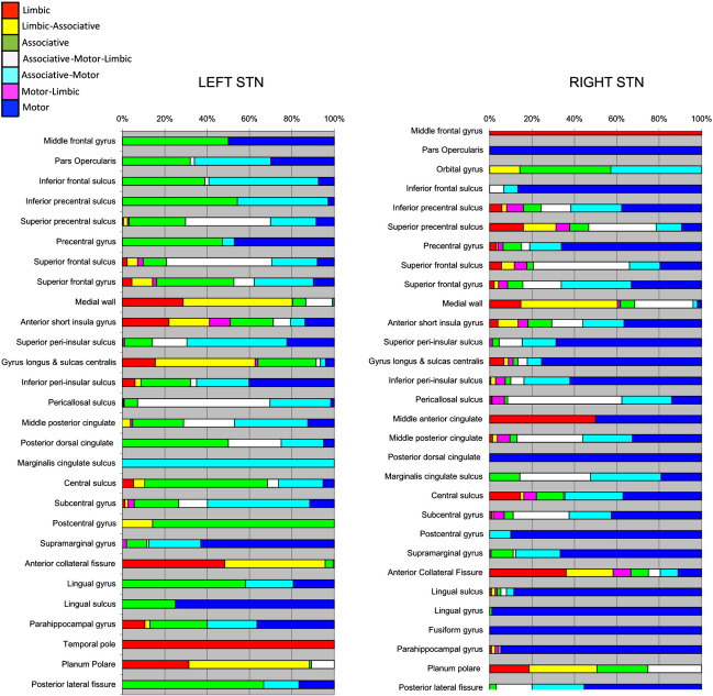
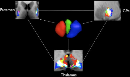
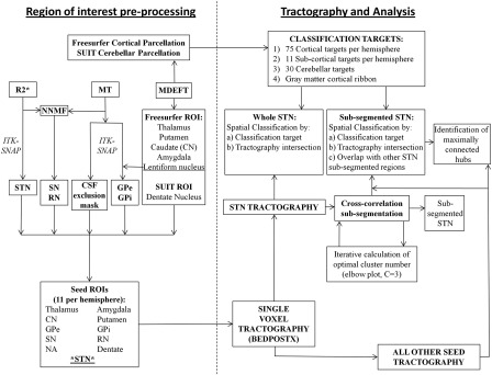
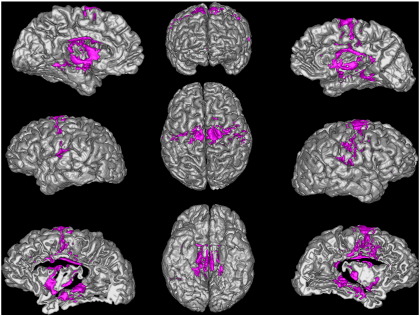
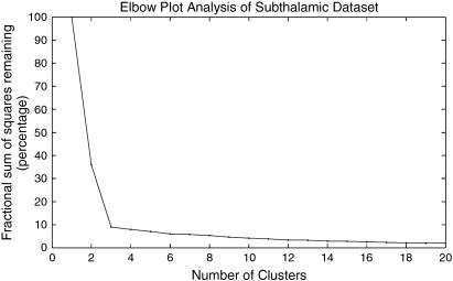
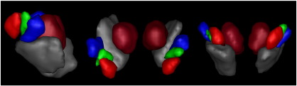
The subthalamic nucleus (STN) is a small, glutamatergic nucleus situated in the diencephalon. A critical component of normal motor function, it has become a key target for deep brain stimulation in the treatment of Parkinson's disease. Animal studies have demonstrated the existence of three functional sub-zones but these have never been shown conclusively in humans. In this work, a data driven method with diffusion weighted imaging demonstrated that three distinct clusters exist within the human STN based on brain connectivity profiles. The STN was successfully sub-parcellated into these regions, demonstrating good correspondence with that described in the animal literature. The local connectivity of each sub-region supported the hypothesis of bilateral limbic, associative and motor regions occupying the anterior, mid and posterior portions of the nucleus respectively. This study is the first to achieve in-vivo, non-invasive anatomical parcellation of the human STN into three anatomical zones within normal diagnostic scan times, which has important future implications for deep brain stimulation surgery.
底丘脑核(STN)是位于间脑的一个小型谷氨酸能核团。作为正常运动功能的关键组成部分,它已成为治疗帕金森病的深部脑刺激的关键靶点。动物研究表明存在三个功能亚区,但这些在人类中从未得到明确证实。在这项工作中,一种基于扩散加权成像的数据分析方法表明,基于脑连接谱,人类 STN 内存在三个不同的簇。STN 被成功地细分到这些区域,与动物文献中描述的结果具有很好的一致性。每个亚区的局部连接支持双侧边缘、联合和运动区域分别占据核的前、中、后部分的假设。这项研究首次在正常诊断扫描时间内,在体内非侵入性地将人类 STN 解剖分区为三个解剖区域,这对深部脑刺激手术具有重要的未来意义。