Department of Biomedical Engineering, University of Wisconsin-Madison, WI 53706, USA.
J Biomech. 2012 Mar 15;45(5):799-804. doi: 10.1016/j.jbiomech.2011.11.020. Epub 2011 Dec 17.
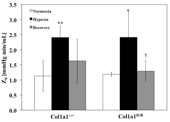
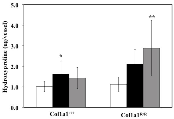
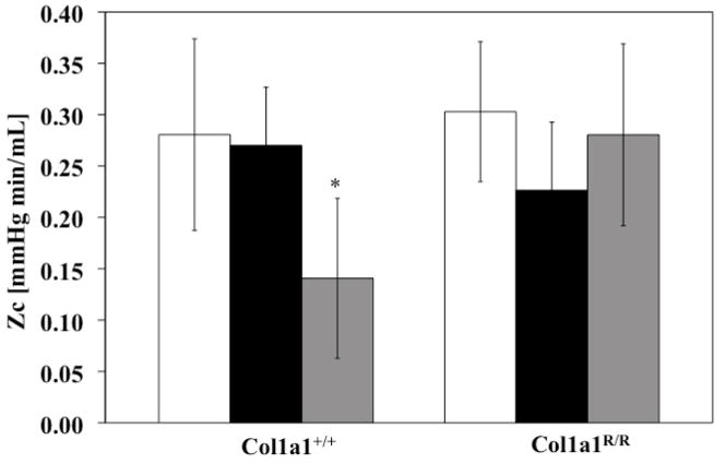
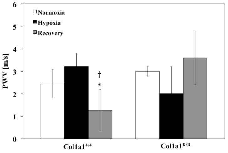
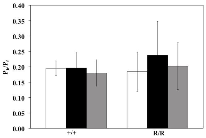
Pulmonary arterial hypertension (PAH) is caused by narrowing and stiffening of the pulmonary arteries that increase pulmonary vascular impedance (PVZ). In particular, small arteries narrow and large arteries stiffen. Large pulmonary artery (PA) stiffness is the best current predictor of mortality from PAH. We have previously shown that collagen accumulation leads to extralobar PA stiffening at high strain (Ooi et al. 2010). We hypothesized that collagen accumulation would increase PVZ, including total pulmonary vascular resistance (Z(0)), characteristic impedance (Z(C)), pulse wave velocity (PWV) and index of global wave reflections (P(b)/P(f)), which contribute to increased right ventricular afterload. We tested this hypothesis by exposing mice unable to degrade type I collagen (Col1a1(R/R)) to 21 days of hypoxia (hypoxia), some of which were allowed to recover for 42 days (recovery). Littermate wild-type mice (Col1a1(+/+)) were used as controls. In response to hypoxia, mean PA pressure (mPAP) increased in both mouse genotypes with no changes in cardiac output (CO) or PA inner diameter (ID); as a consequence, Z(0) (mPAP/CO) increased by ~100% in both genotypes (p<0.05). Contrary to our expectations, Z(C), PWV and P(b)/P(f) did not change. However, with recovery, Z(C) and PWV decreased in the Col1a1(+/+) mice and remained unchanged in the Col1a1(R/R) mice. Z(0) decreased with recovery in both genotypes. Microcomputed tomography measurements of large PAs did not show evidence of stiffness changes as a function of hypoxia exposure or genotype. We conclude that hypoxia-induced PA collagen accumulation does not affect the pulsatile components of pulmonary hemodynamics but that excessive collagen accumulation does prevent normal hemodynamic recovery, which may have important consequences for right ventricular function.
肺动脉高压(PAH)是由肺动脉狭窄和僵硬引起的,这会增加肺血管阻抗(PVZ)。特别是小动脉变窄,大动脉变硬。大动脉僵硬是预测 PAH 死亡率的最佳当前指标。我们之前已经表明,胶原积累会导致高应变下的额外肺段动脉僵硬(Ooi 等人,2010 年)。我们假设胶原积累会增加 PVZ,包括总肺血管阻力(Z(0))、特征阻抗(Z(C))、脉搏波速度(PWV)和全局波反射指数(P(b)/P(f)),这会导致右心室后负荷增加。我们通过使不能降解 I 型胶原的小鼠(Col1a1(R/R))暴露于 21 天的缺氧(缺氧)来测试这一假设,其中一些小鼠允许恢复 42 天(恢复)。同窝野生型小鼠(Col1a1(+/+))用作对照。对缺氧的反应,平均肺动脉压(mPAP)在两种小鼠基因型中均升高,而心输出量(CO)或肺动脉内径(ID)没有变化;因此,两种基因型的 Z(0)(mPAP/CO)均增加了约 100%(p<0.05)。与我们的预期相反,Z(C)、PWV 和 P(b)/P(f) 没有变化。然而,在恢复期间,Col1a1(+/+)小鼠的 Z(C)和 PWV 降低,而 Col1a1(R/R)小鼠的 Z(C)和 PWV 不变。两种基因型的 Z(0)均随恢复而降低。大肺动脉的微计算机断层扫描测量没有显示出随缺氧暴露或基因型变化的僵硬变化的证据。我们得出结论,缺氧诱导的肺动脉胶原积累不会影响肺血流动力学的脉动成分,但过度的胶原积累确实会阻止正常的血流动力学恢复,这可能对右心室功能有重要影响。