Functional and Micro-Magnetic Resonance Imaging Center, Institute of Biomedical Sciences, Academia Sinica, Taipei, Taiwan.
NMR Biomed. 2011 Dec;24(10):1353-60. doi: 10.1002/nbm.1698. Epub 2011 Mar 24.
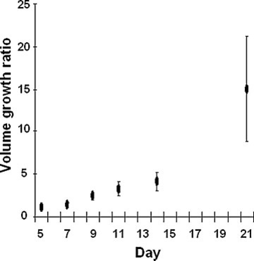
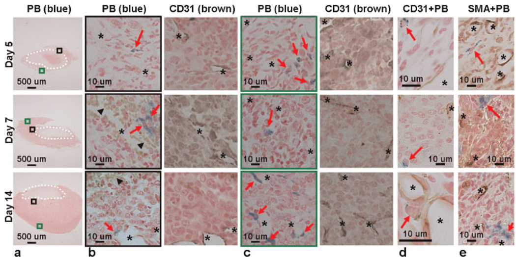
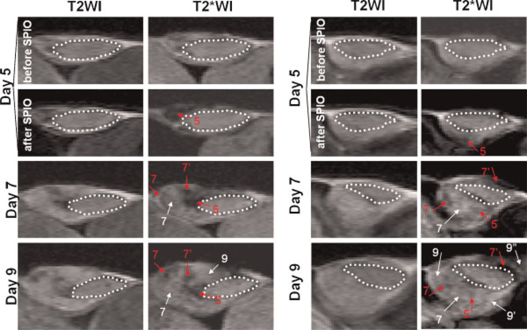
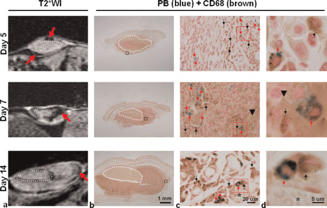
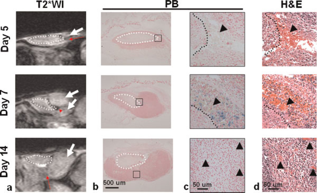
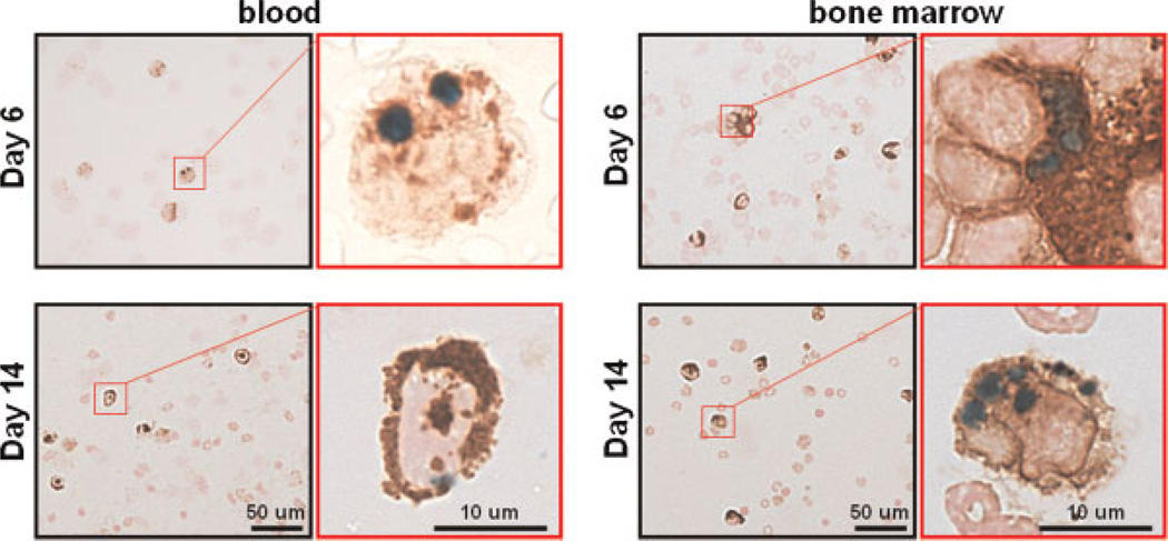
MRI is being used increasingly for the noninvasive longitudinal monitoring of cellular processes in various pathophysiological conditions. Macrophages are the main stromal cells in neoplasms and have been suggested to be the major cell type ingesting superparamagnetic iron oxide (SPIO) nanoparticles. However, no MRI study has described longitudinally the presence of tumor-associated macrophages (TAMs) during tumorigenesis with histological confirmation. To address this, we injected SPIO nanoparticles into the circulation of tumor-bearing mice and used MRI and post-mortem histology to monitor TAMs at different time points. The MRI results demonstrated that TAMs, as hypointense signals, appeared continually with the expansion of the tumor. The histological findings also revealed that SPIO-labeled TAMs tended to deposit closer to the vessel lumen with time prior to rapid tumor growth. The present study demonstrates the potential of using MRI to assess longitudinally TAM accumulation during tumorigenesis, and provides the first in vivo insight into the topographical arrangement of TAMs in relation to the progression of tumors. In vivo monitoring of the presence of TAMs could be useful for the development of tumor treatments that target TAM functions.
磁共振成像(MRI)正被越来越多地用于无创性的、对各种病理生理条件下细胞过程的纵向监测。巨噬细胞是肿瘤中的主要基质细胞,并且被认为是摄取超顺磁性氧化铁(SPIO)纳米颗粒的主要细胞类型。然而,目前还没有 MRI 研究描述了在肿瘤发生过程中具有组织学确认的肿瘤相关巨噬细胞(TAMs)的存在。为了解决这个问题,我们将 SPIO 纳米颗粒注入荷瘤小鼠的循环系统中,并使用 MRI 和死后组织学来监测不同时间点的 TAMs。MRI 结果表明,TAMs 作为低信号出现,随着肿瘤的扩张而持续存在。组织学发现还表明,SPIO 标记的 TAMs 倾向于在肿瘤快速生长之前,随着时间的推移更靠近血管腔沉积。本研究表明了使用 MRI 来评估肿瘤发生过程中 TAM 积累的潜力,并首次提供了关于 TAMs 在肿瘤进展过程中的拓扑排列的体内见解。TAMs 存在的体内监测可能有助于开发针对 TAM 功能的肿瘤治疗方法。