Clarendon Laboratory, Department of Physics, University of Oxford, Oxford, UK.
Nat Struct Mol Biol. 2012 Jan 8;19(2):158-63. doi: 10.1038/nsmb.2208.
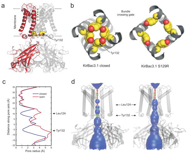
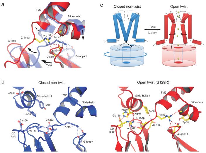
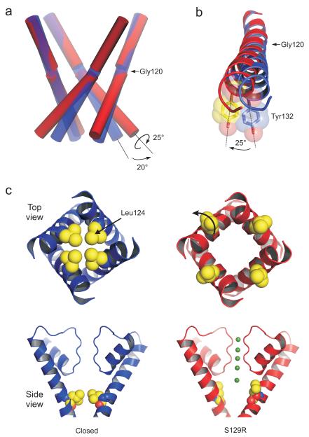
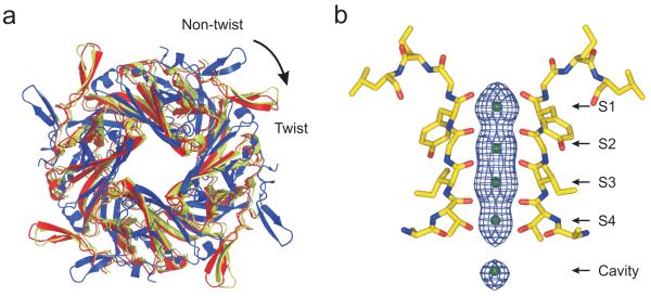
KirBac channels are prokaryotic homologs of mammalian inwardly rectifying (Kir) potassium channels, and recent crystal structures of both Kir and KirBac channels have provided major insight into their unique structural architecture. However, all of the available structures are closed at the helix bundle crossing, and therefore the structural mechanisms that control opening of their primary activation gate remain unknown. In this study, we engineered the inner pore-lining helix (TM2) of KirBac3.1 to trap the bundle crossing in an apparently open conformation and determined the crystal structure of this mutant channel to 3.05 Å resolution. Contrary to previous speculation, this new structure suggests a mechanistic model in which rotational 'twist' of the cytoplasmic domain is coupled to opening of the bundle-crossing gate through a network of inter- and intrasubunit interactions that involve the TM2 C-linker, slide helix, G-loop and the CD loop.
KirBac 通道是哺乳动物内向整流 (Kir) 钾通道的原核同源物,最近 Kir 和 KirBac 通道的晶体结构为它们独特的结构架构提供了重要的见解。然而,所有可用的结构在螺旋束交叉处都是关闭的,因此控制其主要激活门打开的结构机制仍然未知。在这项研究中,我们对 KirBac3.1 的内孔衬里螺旋 (TM2) 进行了工程改造,以将束交叉捕获在明显开放的构象中,并确定了该突变通道的晶体结构,分辨率为 3.05 Å。与之前的推测相反,这个新结构提出了一个机械模型,其中细胞质结构域的旋转“扭曲”通过涉及 TM2 C 接头、滑动螺旋、G 环和 CD 环的相互和亚基间相互作用与束交叉门的打开相耦合。