Department of Infectious Diseases, Faculty of Medicine, University of Gaziantep, Gaziantep, Turkey.
J Infect Chemother. 2012 Oct;18(5):767-70. doi: 10.1007/s10156-011-0365-4. Epub 2012 Jan 11.
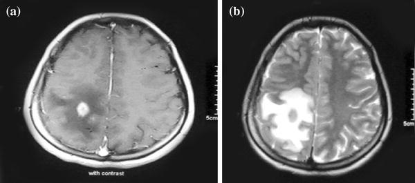
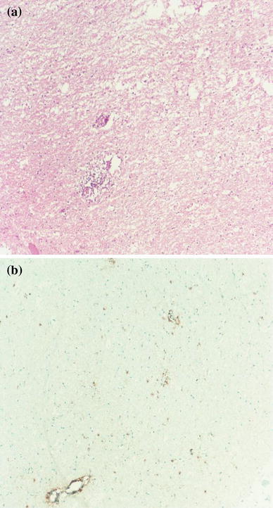
Among the diverse presentations of neurobrucellosis, solitary intracranial mass lesions are extremely rare. To the best of our knowledge, we describe here the second case of neurobrucellosis mimicking a cerebral tumor caused by Brucella melitensis. The mass lesion was clinically and radiologically indistinguishable from a brain tumor. The diagnosis was established by isolating Brucella melitensis in a blood culture and a positive Wright's agglutination test on the cerebrospinal fluid at 1:320 titers. Paraffin sections of the cerebral mass showed nongranulomatous encephalitis. We suggest that patients with an isolated intraparenchymal mass lesion with nongranulomatous encephalitis should also be studied for brucellosis in endemic areas.
在神经布鲁氏菌病的多种表现中,孤立性颅内肿块病变极为罕见。据我们所知,我们在这里描述了第二个由布鲁氏菌引起的神经布鲁氏菌病模拟脑瘤的病例。该肿块病变在临床和影像学上与脑瘤无法区分。通过在血液培养中分离出布鲁氏菌melitensis 以及脑脊液中的 Wright 凝集试验滴度为 1:320 阳性,确诊了该诊断。脑肿块的石蜡切片显示非化脓性脑炎。我们建议,在布鲁氏菌病流行地区,对于伴有非化脓性脑炎的孤立性脑实质肿块病变患者,也应进行布鲁氏菌病研究。