Department of Neurosurgery, Frenchay Hospital, Bristol, BS16 1LE, UK.
Fluids Barriers CNS. 2012 Jan 20;9(1):2. doi: 10.1186/2045-8118-9-2.
Convection-enhanced delivery (CED), a direct method for drug delivery to the brain through intraparenchymal microcatheters, is a promising strategy for intracerebral pharmacological therapy. By establishing a pressure gradient at the tip of the catheter, drugs can be delivered in uniform concentration throughout a large volume of interstitial fluid. However, the variables affecting perivascular distribution of drugs delivered by CED are not fully understood. The aim of this study was to determine whether the perivascular distribution of solutes delivered by CED into the striatum of rats is affected by the molecular weight of the infused agent, by co-infusion of vasodilator, alteration of infusion rates or use of a ramping regime. We also wanted to make a preliminary comparison of the distribution of solutes with that of nanoparticles.
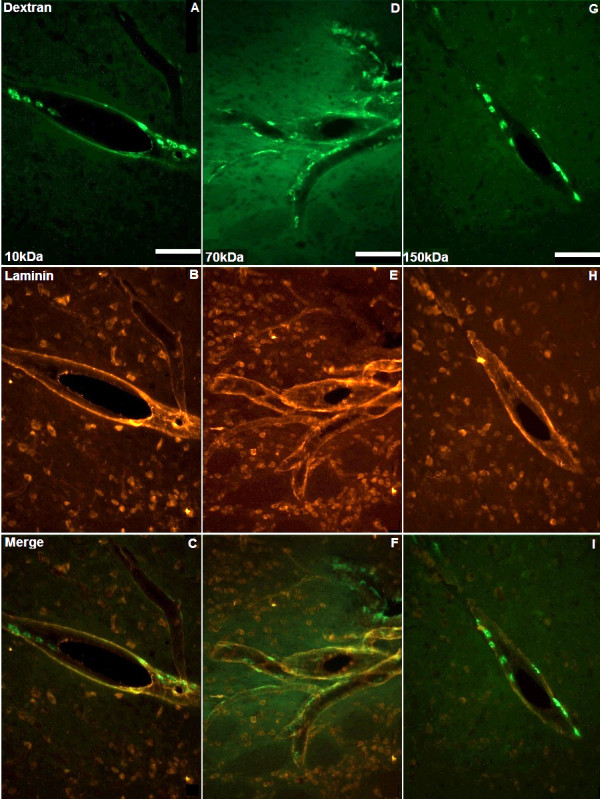
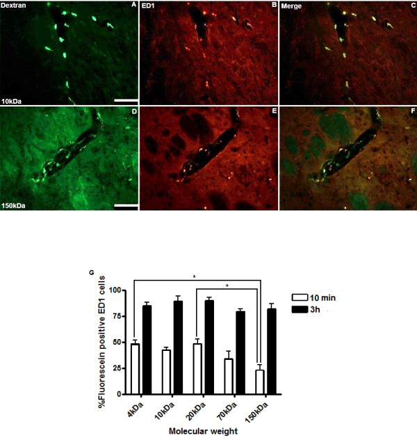
We analysed the perivascular distribution of 4, 10, 20, 70, 150 kDa fluorescein-labelled dextran and fluorescent nanoparticles at 10 min and 3 h following CED into rat striatum. We investigated the effect of local vasodilatation, slow infusion rates and ramping on the perivascular distribution of solutes. Co-localisation with perivascular basement membranes and vascular endothelial cells was identified by immunohistochemistry. The uptake of infusates by perivascular macrophages was quantified using stereological methods.
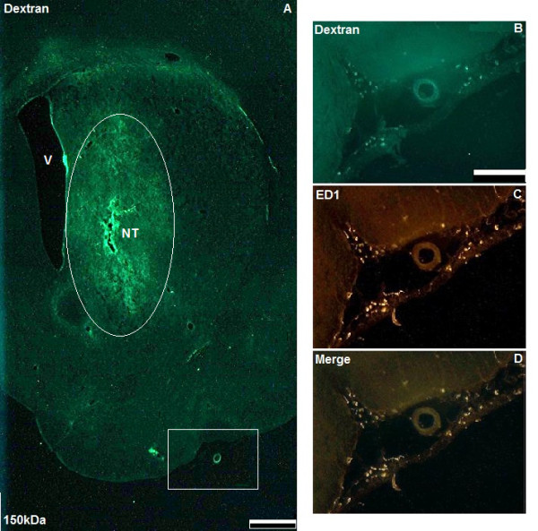
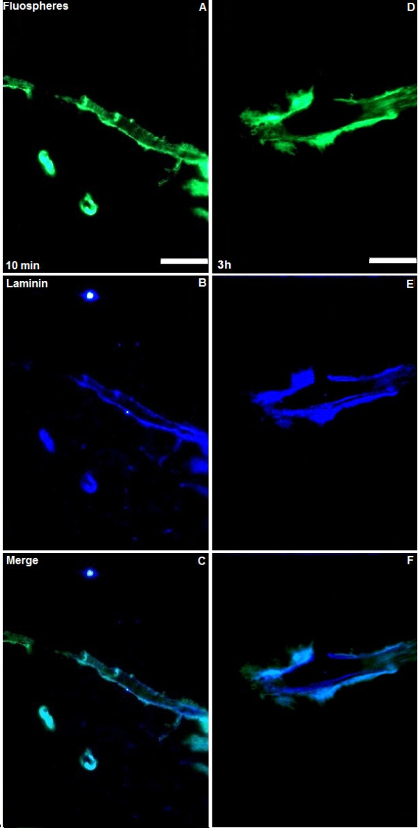
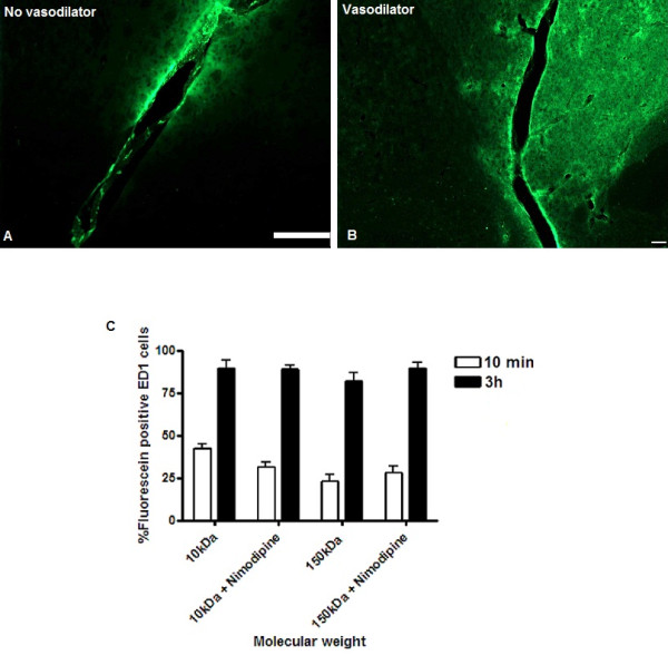
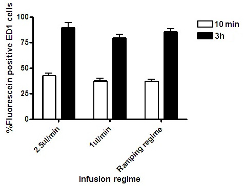
Widespread perivascular distribution and macrophage uptake of fluorescein-labelled dextran was visible 10 min after cessation of CED irrespective of molecular weight. However, a significantly higher proportion of perivascular macrophages had taken up 4, 10 and 20 kDa fluorescein-labelled dextran than 150 kDa dextran (p < 0.05, ANOVA). Co-infusion with vasodilator, slow infusion rates and use of a ramping regime did not alter the perivascular distribution. CED of fluorescent nanoparticles indicated that particles co-localise with perivascular basement membranes throughout the striatum but, unlike soluble dextrans, are not taken up by perivascular macrophages after 3 h.
This study suggests that widespread perivascular distribution and interaction with perivascular macrophages is likely to be an inevitable consequence of CED of solutes. The potential consequences of perivascular distribution of therapeutic agents, and in particular cytotoxic chemotherapies, delivered by CED must be carefully considered to ensure safe and effective translation to clinical trials.
通过脑实质微导管进行的增强型传递(CED)是一种将药物直接递送至大脑的方法,是脑内药物治疗的一种有前途的策略。通过在导管尖端建立压力梯度,可以将药物均匀地递送到大体积的间质液中。然而,影响通过 CED 递送至血管周围的药物分布的变量尚未完全了解。本研究旨在确定通过 CED 递送至大鼠纹状体的溶质的血管周围分布是否受输注剂分子量、共输注血管扩张剂、改变输注率或使用斜坡方案的影响。我们还想初步比较溶质和纳米颗粒的分布。
我们分析了 4、10、20、70、150 kDa 荧光素标记的葡聚糖和荧光纳米颗粒在 CED 后 10 分钟和 3 小时在大鼠纹状体中的血管周围分布。我们研究了局部血管扩张、缓慢输注率和斜坡对溶质血管周围分布的影响。通过免疫组织化学鉴定与血管周围基膜和血管内皮细胞的共定位。使用体视学法定量测量血管周围巨噬细胞对灌注液的摄取。
CED 停止后 10 分钟,无论分子量如何,均可观察到广泛的血管周围分布和荧光素标记的葡聚糖被血管周围巨噬细胞摄取。然而,与 150 kDa 葡聚糖相比,4、10 和 20 kDa 荧光素标记的葡聚糖被更多的血管周围巨噬细胞摄取(p < 0.05,方差分析)。共输注血管扩张剂、缓慢输注率和使用斜坡方案并未改变血管周围的分布。荧光纳米颗粒的 CED 表明,颗粒与整个纹状体的血管周围基膜共定位,但与可溶葡聚糖不同,3 小时后不会被血管周围巨噬细胞摄取。
本研究表明,广泛的血管周围分布和与血管周围巨噬细胞的相互作用可能是 CED 输送溶质不可避免的结果。必须仔细考虑通过 CED 输送的治疗剂,特别是细胞毒性化疗药物的血管周围分布的潜在后果,以确保安全有效地转化为临床试验。