• 文献检索
  • 文档翻译
  • 深度研究
  • 学术资讯
  • Suppr Zotero 插件Zotero 插件
  • 邀请有礼
  • 套餐&价格
  • 历史记录
应用&插件
Suppr Zotero 插件Zotero 插件浏览器插件Mac 客户端Windows 客户端微信小程序
定价
高级版会员购买积分包购买API积分包
服务
文献检索文档翻译深度研究API 文档MCP 服务
关于我们
关于 Suppr公司介绍联系我们用户协议隐私条款
关注我们

Suppr 超能文献

核心技术专利:CN118964589B侵权必究
粤ICP备2023148730 号-1Suppr @ 2026

文献检索

告别复杂PubMed语法,用中文像聊天一样搜索,搜遍4000万医学文献。AI智能推荐,让科研检索更轻松。

立即免费搜索

文件翻译

保留排版,准确专业,支持PDF/Word/PPT等文件格式,支持 12+语言互译。

免费翻译文档

深度研究

AI帮你快速写综述,25分钟生成高质量综述,智能提取关键信息,辅助科研写作。

立即免费体验

SD-OCT数据集的自动拼接

Automatic montage of SD-OCT data sets.

作者信息

Li Ying, Gregori Giovanni, Lam Byron L, Rosenfeld Philip J

机构信息

Department of Ophthalmology, Bascom Palmer Eye Institute, University of Miami Miller School of Medicine, Miami, Florida 33136, USA.

出版信息

Opt Express. 2011 Dec 19;19(27):26239-48. doi: 10.1364/OE.19.026239.

DOI:10.1364/OE.19.026239
PMID:22274209
原文链接:https://pmc.ncbi.nlm.nih.gov/articles/PMC3500106/
Abstract

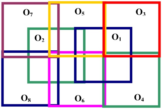
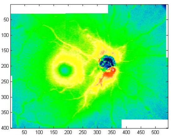
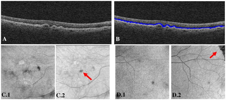
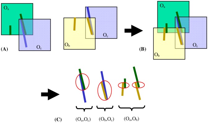
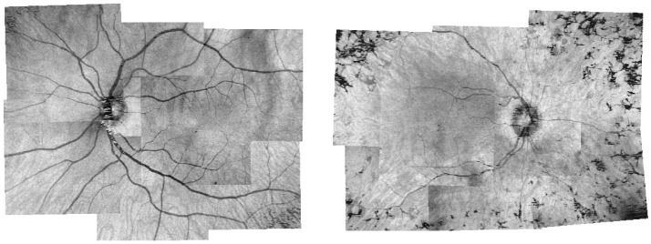
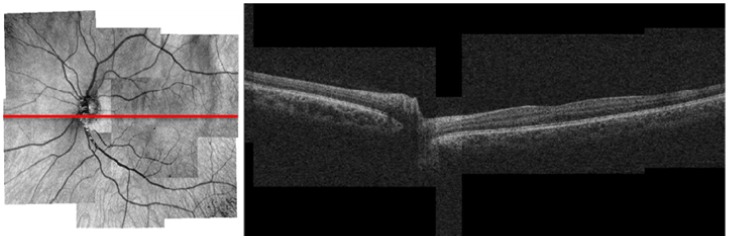
This paper proposes an automatic algorithm for the montage of OCT data sets, which produces a composite 3D OCT image over a large field of view out of several separate, partially overlapping OCT data sets. First the OCT fundus images (OFIs) are registered, using blood vessel ridges as the feature of interest and a two step iterative procedure to minimize the distance between all matching point pairs over the set of OFIs. Then the OCT data sets are merged to form a full 3D montage using cross-correlation. The algorithm was tested using an imaging protocol consisting of 8 OCT images for each eye, overlapping to cover a total retinal region of approximately 50x35 degrees. The results for 3 normal eyes and 3 eyes with retinal degeneration are analyzed, showing registration errors of 1.5±0.3 and 2.0±0.8 pixels respectively.

摘要

本文提出了一种用于OCT数据集拼接的自动算法,该算法可从几个单独的、部分重叠的OCT数据集中生成一个大视野的复合3D OCT图像。首先,以血管嵴作为感兴趣特征,并采用两步迭代程序来最小化OCT眼底图像(OFI)集合中所有匹配点对之间的距离,从而对OFI进行配准。然后,使用互相关将OCT数据集合并以形成完整的3D拼接图像。该算法通过一种成像协议进行测试,该协议为每只眼睛采集8张OCT图像,这些图像相互重叠以覆盖大约50×35度的整个视网膜区域。分析了3只正常眼睛和3只患有视网膜变性眼睛的结果,结果显示配准误差分别为1.5±0.3像素和2.0±0.8像素。

https://cdn.ncbi.nlm.nih.gov/pmc/blobs/633f/3500106/c51289b54d98/oe-19-27-26239-g006.jpg
https://cdn.ncbi.nlm.nih.gov/pmc/blobs/633f/3500106/d41971b13e90/oe-19-27-26239-g001.jpg
https://cdn.ncbi.nlm.nih.gov/pmc/blobs/633f/3500106/1cda47d00aa5/oe-19-27-26239-g002.jpg
https://cdn.ncbi.nlm.nih.gov/pmc/blobs/633f/3500106/e9c99c6b3288/oe-19-27-26239-g003.jpg
https://cdn.ncbi.nlm.nih.gov/pmc/blobs/633f/3500106/be37aa29151f/oe-19-27-26239-g004.jpg
https://cdn.ncbi.nlm.nih.gov/pmc/blobs/633f/3500106/4752972fe460/oe-19-27-26239-g005.jpg
https://cdn.ncbi.nlm.nih.gov/pmc/blobs/633f/3500106/c51289b54d98/oe-19-27-26239-g006.jpg

相似文献

1
Automatic montage of SD-OCT data sets.SD-OCT数据集的自动拼接
Opt Express. 2011 Dec 19;19(27):26239-48. doi: 10.1364/OE.19.026239.
2
Exact surface registration of retinal surfaces from 3-D optical coherence tomography images.从三维光学相干断层扫描图像中实现视网膜表面的精确表面配准。
IEEE Trans Biomed Eng. 2015 Feb;62(2):609-17. doi: 10.1109/TBME.2014.2361778. Epub 2014 Oct 8.
3
Motion artefact correction in retinal optical coherence tomography using local symmetry.利用局部对称性校正视网膜光学相干断层扫描中的运动伪影
Med Image Comput Comput Assist Interv. 2014;17(Pt 2):130-7. doi: 10.1007/978-3-319-10470-6_17.
4
Improving image segmentation performance and quantitative analysis via a computer-aided grading methodology for optical coherence tomography retinal image analysis.通过光学相干断层扫描视网膜图像分析的计算机辅助分级方法提高图像分割性能和定量分析。
J Biomed Opt. 2010 Jul-Aug;15(4):046015. doi: 10.1117/1.3470116.
5
A generative model for OCT retinal layer segmentation by integrating graph-based multi-surface searching and image registration.一种通过整合基于图的多表面搜索和图像配准的光学相干断层扫描(OCT)视网膜层分割生成模型。
Med Image Comput Comput Assist Interv. 2013;16(Pt 1):428-35. doi: 10.1007/978-3-642-40811-3_54.
6
Enhancing the signal-to-noise ratio in ophthalmic optical coherence tomography by image registration--method and clinical examples.通过图像配准提高眼科光学相干断层扫描中的信噪比——方法及临床实例
J Biomed Opt. 2007 Jul-Aug;12(4):041208. doi: 10.1117/1.2772879.
7
User-guided segmentation for volumetric retinal optical coherence tomography images.用于视网膜光学相干断层扫描容积图像的用户引导分割
J Biomed Opt. 2014 Aug;19(8):086020. doi: 10.1117/1.JBO.19.8.086020.
8
[Comparison of spectral- and time-domain OCT in regard of the automatic detection of central retinal thickness].[光谱域光学相干断层扫描与时域光学相干断层扫描在自动检测视网膜中央厚度方面的比较]
Klin Monbl Augenheilkd. 2009 Feb;226(2):115-9. doi: 10.1055/s-2008-1027913. Epub 2009 Feb 10.
9
Objective and expert-independent validation of retinal image registration algorithms by a projective imaging distortion model.利用投影成像失真模型对视网膜图像配准算法进行目标和专家独立验证。
Med Image Anal. 2010 Aug;14(4):539-49. doi: 10.1016/j.media.2010.04.001. Epub 2010 Apr 28.
10
Automated drusen segmentation and quantification in SD-OCT images.SD-OCT 图像中的自动 drusen 分割和定量。
Med Image Anal. 2013 Dec;17(8):1058-72. doi: 10.1016/j.media.2013.06.003. Epub 2013 Jul 2.

引用本文的文献

1
Adaptive point-scan imaging beyond the frame rate-resolution limit with scene-reactive scan trajectories.采用场景反应扫描轨迹突破帧率分辨率限制的自适应点扫描成像。
Optica. 2022 Nov 20;9(11):1276-1288. doi: 10.1364/optica.472562.
2
Extending field-of-view of retinal imaging by optical coherence tomography using convolutional Lissajous and slow scan patterns.利用卷积李萨如和慢扫描模式通过光学相干断层扫描扩展视网膜成像的视野
Biomed Opt Express. 2022 Sep 9;13(10):5212-5230. doi: 10.1364/BOE.467563. eCollection 2022 Oct 1.
3
Generating large field of view en-face projection images from intra-acquisition motion compensated volumetric optical coherence tomography data.

本文引用的文献

1
Speckle in optical coherence tomography.光学相干断层扫描中的散斑
J Biomed Opt. 1999 Jan;4(1):95-105. doi: 10.1117/1.429925.
2
Increasing the field-of-view of dynamic cardiac OCT via post-acquisition mosaicing without affecting frame-rate or spatial resolution.通过采集后拼接增加动态心脏光学相干断层扫描的视野,同时不影响帧率或空间分辨率。
Biomed Opt Express. 2011 Sep 1;2(9):2614-22. doi: 10.1364/BOE.2.002614. Epub 2011 Aug 16.
3
Spectral domain optical coherence tomography imaging of drusen in nonexudative age-related macular degeneration.
从采集内运动补偿的体层光学相干断层扫描数据生成大视野正面投影图像。
Biomed Opt Express. 2020 Nov 4;11(12):6881-6904. doi: 10.1364/BOE.404738. eCollection 2020 Dec 1.
4
A Deep-Learning Approach for Automated OCT En-Face Retinal Vessel Segmentation in Cases of Optic Disc Swelling Using Multiple En-Face Images as Input.一种基于深度学习的方法,用于使用多张 OCT 眼前节图像作为输入,对视盘肿胀病例进行 OCT 自动眼前节视网膜血管分割。
Transl Vis Sci Technol. 2020 Mar 24;9(2):17. doi: 10.1167/tvst.9.2.17. eCollection 2020 Mar.
5
High-resolution wide-field OCT angiography with a self-navigation method to correct microsaccades and blinks.采用自导航方法校正微扫视和眨眼的高分辨率宽视野光学相干断层扫描血管造影术。
Biomed Opt Express. 2020 May 21;11(6):3234-3245. doi: 10.1364/BOE.390430. eCollection 2020 Jun 1.
6
Peripheral optical coherence tomography montage guided successful management of retinal detachment with sub retinal bands.周边光学相干断层扫描拼接图引导下成功治疗伴有视网膜下条索的视网膜脱离。
Saudi J Ophthalmol. 2017 Oct-Dec;31(4):282-284. doi: 10.1016/j.sjopt.2017.04.001. Epub 2017 Apr 9.
7
Involuntary eye motion correction in retinal optical coherence tomography: Hardware or software solution?视网膜光学相干断层扫描中的非自主眼动校正:硬件还是软件解决方案?
Med Image Anal. 2017 Apr;37:129-145. doi: 10.1016/j.media.2017.02.002. Epub 2017 Feb 4.
8
A Mosaicking Approach for In Vivo Thickness Mapping of the Human Tympanic Membrane Using Low Coherence Interferometry.一种使用低相干干涉测量法对人鼓膜进行体内厚度映射的拼接方法。
J Assoc Res Otolaryngol. 2016 Oct;17(5):403-16. doi: 10.1007/s10162-016-0576-6. Epub 2016 Jul 25.
9
Tissue thickness calculation in ocular optical coherence tomography.眼部光学相干断层扫描中的组织厚度计算。
Biomed Opt Express. 2016 Jan 21;7(2):629-45. doi: 10.1364/BOE.7.000629. eCollection 2016 Feb 1.
10
State-of-the-art in retinal optical coherence tomography image analysis.视网膜光学相干断层扫描图像分析的最新技术。
Quant Imaging Med Surg. 2015 Aug;5(4):603-17. doi: 10.3978/j.issn.2223-4292.2015.07.02.
非渗出性年龄相关性黄斑变性中玻璃膜疣的光谱域光学相干断层成像。
Ophthalmology. 2011 Jul;118(7):1373-9. doi: 10.1016/j.ophtha.2010.11.013. Epub 2011 Mar 9.
4
Megahertz OCT for ultrawide-field retinal imaging with a 1050 nm Fourier domain mode-locked laser.采用1050纳米傅里叶域锁模激光器的兆赫兹光学相干断层扫描技术用于超广角视网膜成像
Opt Express. 2011 Feb 14;19(4):3044-62. doi: 10.1364/OE.19.003044.
5
Registration of OCT fundus images with color fundus photographs based on blood vessel ridges.基于血管嵴的光学相干断层扫描眼底图像与彩色眼底照片配准
Opt Express. 2011 Jan 3;19(1):7-16. doi: 10.1364/OE.19.000007.
6
Salient feature region: a new method for retinal image registration.显著特征区域:一种用于视网膜图像配准的新方法。
IEEE Trans Inf Technol Biomed. 2011 Mar;15(2):221-32. doi: 10.1109/TITB.2010.2091145. Epub 2010 Dec 6.
7
Spectral domain optical coherence tomography imaging of dry age-related macular degeneration.干性年龄相关性黄斑变性的光谱域光学相干断层扫描成像
Ophthalmic Surg Lasers Imaging. 2010 Nov-Dec;41 Suppl:S6-S14. doi: 10.3928/15428877-20101031-19.
8
Automated layer segmentation of macular OCT images using dual-scale gradient information.利用双尺度梯度信息对黄斑OCT图像进行自动层分割。
Opt Express. 2010 Sep 27;18(20):21293-307. doi: 10.1364/OE.18.021293.
9
Ultrahigh speed 1050nm swept source/Fourier domain OCT retinal and anterior segment imaging at 100,000 to 400,000 axial scans per second.超高速1050纳米扫频光源/傅里叶域光学相干断层扫描技术,每秒可进行100000至400000次轴向扫描,用于视网膜和眼前节成像。
Opt Express. 2010 Sep 13;18(19):20029-48. doi: 10.1364/OE.18.020029.
10
Automated segmentation of 3-D spectral OCT retinal blood vessels by neural canal opening false positive suppression.通过神经管开口假阳性抑制实现三维光谱光学相干断层扫描视网膜血管的自动分割。
Med Image Comput Comput Assist Interv. 2010;13(Pt 3):33-40. doi: 10.1007/978-3-642-15711-0_5.