Pierro Luisa, Zampedri Elena, Milani Paolo, Gagliardi Marco, Isola Vincenzo, Pece Alfredo
Department of Ophthalmology, University Vita-Salute, Scientific Institute San Raffaele, Milano, Italy.
Clin Ophthalmol. 2012;6:219-23. doi: 10.2147/OPTH.S27656. Epub 2012 Feb 9.
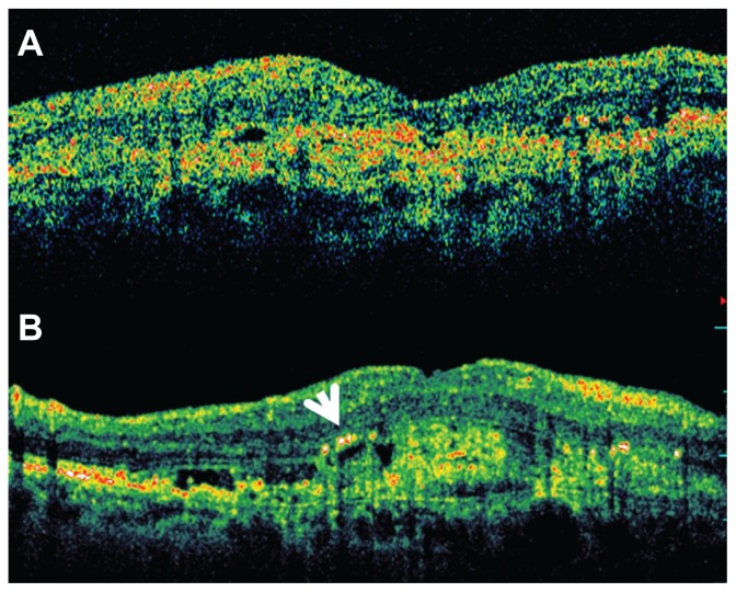
The aim of this study was to compare the agreement between spectral domain optical coherence tomography (SD OCT) and time domain stratus OCT (TD OCT) in evaluating macular morphology alterations in wet age-related macular degeneration (AMD).
This retrospective study was performed on 77 eyes of 77 patients with primary or recurring subfoveal choroidal neovascularization secondary to AMD. All patients underwent OCT examination using Zeiss Stratus OCT 3 (Carl Zeiss Meditec Inc, Dublin, CA) and Opko OTI Spectral SLO/OCT (Ophthalmic Technologies Inc, Toronto, Canada). In all radial line scans, the presence of intraretinal edema (IRE), serous pigment epithelium detachment (sPED), neurosensory serous retinal detachment (NSRD), epiretinal membrane (EM), inner limiting membrane thickening (ILMT), and hard exudates (HE) were evaluated. The degree of matching was quantified by Kappa measure of agreement.
THE PERCENTAGE DISTRIBUTION OF TD OCT FINDINGS VERSUS SD OCT FINDINGS WAS: IRE 36.3% versus 77.9%, sPED 57.1% versus 85.7%, NSRD 38.9% versus 53.2%, EM 10.5% versus 26.3%, ILMT 3.8% versus 32.4%, and HE 6.4% versus 54.5%. The agreement was as follows: sPED: kappa value 0.15; NSRD: kappa value 0.61; IRE: kappa value 0.18; EM: kappa value 0.41; ILMT: kappa value 0.02; HE: kappa value 0.06.
The agreement in the evaluation of macular lesions between the two techniques is poor and depends on the lesion considered. SD OCT allows better detection of the alterations typically related to choroidal neovascularization such as IRE, PED, ILM thickening, and HE. Consequently its use should be strongly considered in patients with wet AMD.
本研究旨在比较频域光学相干断层扫描(SD OCT)和时域Stratus OCT(TD OCT)在评估湿性年龄相关性黄斑变性(AMD)黄斑形态改变方面的一致性。
对77例原发性或复发性继发于AMD的黄斑下脉络膜新生血管患者的77只眼进行了这项回顾性研究。所有患者均使用蔡司Stratus OCT 3(卡尔蔡司医疗技术公司,加利福尼亚州都柏林)和Opko OTI Spectral SLO/OCT(眼科技术公司,加拿大多伦多)进行了OCT检查。在所有径向线扫描中,评估视网膜内水肿(IRE)、浆液性色素上皮脱离(sPED)、神经感觉性浆液性视网膜脱离(NSRD)、视网膜前膜(EM)、内界膜增厚(ILMT)和硬性渗出(HE)的存在情况。通过Kappa一致性测量对匹配程度进行量化。
TD OCT结果与SD OCT结果的百分比分布如下:IRE为36.3%对77.9%,sPED为57.1%对85.7%,NSRD为38.9%对53.2%,EM为10.5%对26.3%,ILMT为3.8%对32.4%,HE为6.4%对54.5%。一致性如下:sPED:kappa值0.15;NSRD:kappa值0.61;IRE:kappa值0.18;EM:kappa值0.41;ILMT:kappa值0.02;HE:kappa值0.06。
两种技术在评估黄斑病变方面的一致性较差,且取决于所考虑的病变。SD OCT能更好地检测出通常与脉络膜新生血管相关的改变,如IRE、PED、ILM增厚和HE。因此,对于湿性AMD患者,应强烈考虑使用SD OCT。