Duarte Ricardo, Cisneros Silvia, Fernandez Gabriel, Castellon Daniel, Cattani Cesar, A Melo Cíntia, Apocada Asier
Insights Imaging. 2010 Sep;1(4):223-231. doi: 10.1007/s13244-010-0035-6. Epub 2010 Jul 30.
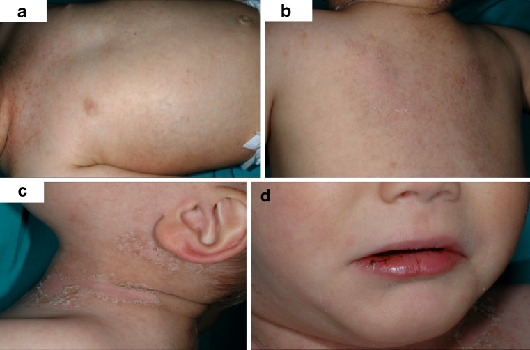
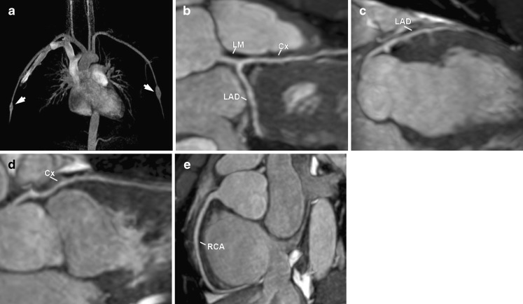
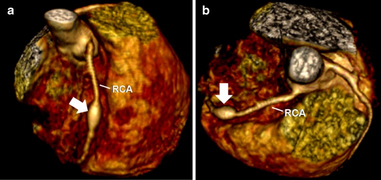
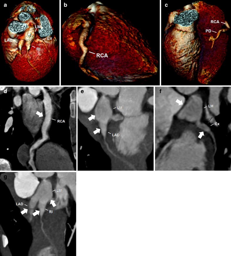
Kawasaki disease (KD) is an acute systemic vasculitis that is currently the leading cause of acquired heart disease in childhood in the United States. Cardiovascular complications are the major cause of morbidity, are responsible for virtually all deaths from KD and should be evaluated as soon as possible after the acute phase to establish the baseline status, in order to predict disease progression and determine adequate treatment. In selected patients, electrocardiography (ECG)-gated cardiac computed tomography (CT) and magnetic resonance (MR) imaging are valuable non-invasive techniques that can be used to help diagnose the cardiovascular complications associated with KD. In this article, we review the epidemiology, aetiology and pathogenesis, histopathology, clinical features, cardiovascular complications and imaging, focusing on the role of cardiac CT and MR on the initial assessment and follow-up of the cardiovascular complications of KD.
川崎病(KD)是一种急性全身性血管炎,目前是美国儿童后天性心脏病的主要病因。心血管并发症是发病的主要原因,几乎是KD所有死亡的原因,急性期后应尽快进行评估以确定基线状态,以便预测疾病进展并确定适当的治疗方法。对于部分患者,心电图(ECG)门控心脏计算机断层扫描(CT)和磁共振(MR)成像都是有价值的非侵入性技术,可用于帮助诊断与KD相关的心血管并发症。在本文中,我们回顾了KD的流行病学、病因学和发病机制、组织病理学、临床特征、心血管并发症及影像学,重点关注心脏CT和MR在KD心血管并发症的初始评估和随访中的作用。