• 文献检索
  • 文档翻译
  • 深度研究
  • 学术资讯
  • Suppr Zotero 插件Zotero 插件
  • 邀请有礼
  • 套餐&价格
  • 历史记录
应用&插件
Suppr Zotero 插件Zotero 插件浏览器插件Mac 客户端Windows 客户端微信小程序
定价
高级版会员购买积分包购买API积分包
服务
文献检索文档翻译深度研究API 文档MCP 服务
关于我们
关于 Suppr公司介绍联系我们用户协议隐私条款
关注我们

Suppr 超能文献

核心技术专利:CN118964589B侵权必究
粤ICP备2023148730 号-1Suppr @ 2026

文献检索

告别复杂PubMed语法,用中文像聊天一样搜索,搜遍4000万医学文献。AI智能推荐,让科研检索更轻松。

立即免费搜索

文件翻译

保留排版,准确专业,支持PDF/Word/PPT等文件格式,支持 12+语言互译。

免费翻译文档

深度研究

AI帮你快速写综述,25分钟生成高质量综述,智能提取关键信息,辅助科研写作。

立即免费体验

肝硬化的磁共振成像:传统方法与新方法

MR imaging in liver cirrhosis: classical and new approaches.

作者信息

Martí-Bonmatí Luis, Delgado Fructuoso

出版信息

Insights Imaging. 2010 Sep;1(4):233-244. doi: 10.1007/s13244-010-0034-7. Epub 2010 Aug 5.

DOI:10.1007/s13244-010-0034-7
PMID:22347919
原文链接:https://pmc.ncbi.nlm.nih.gov/articles/PMC3259325/
Abstract

OBJECTIVE

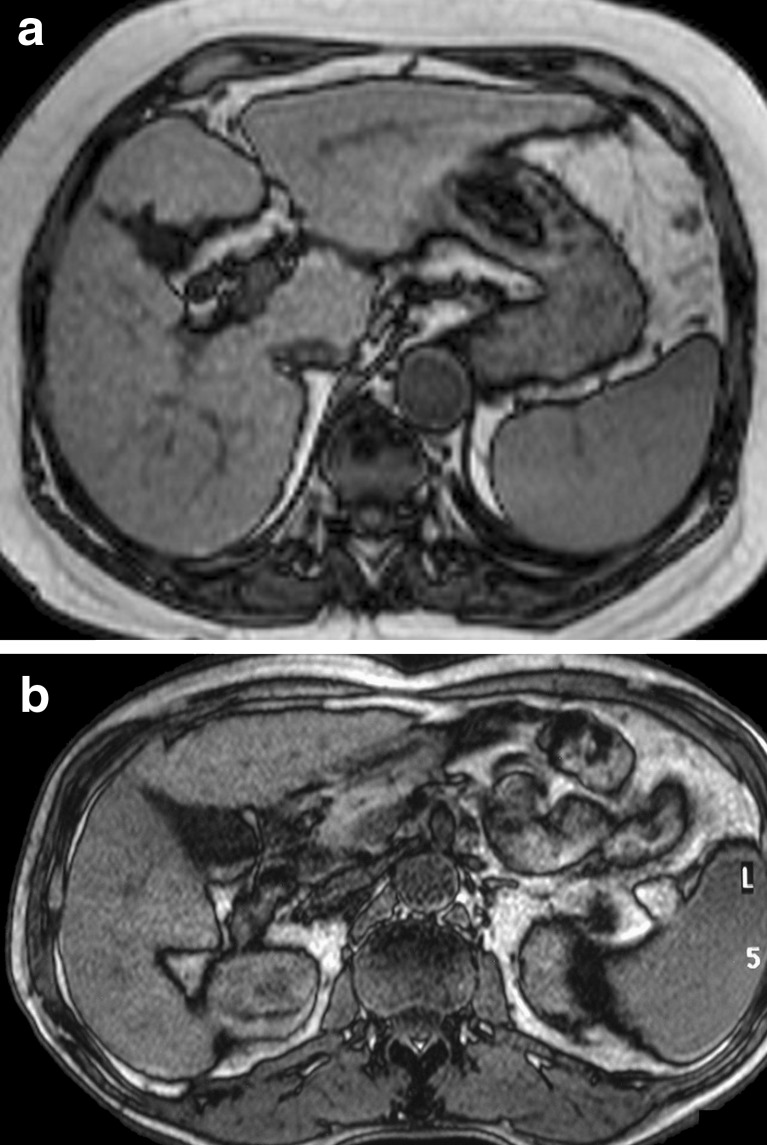
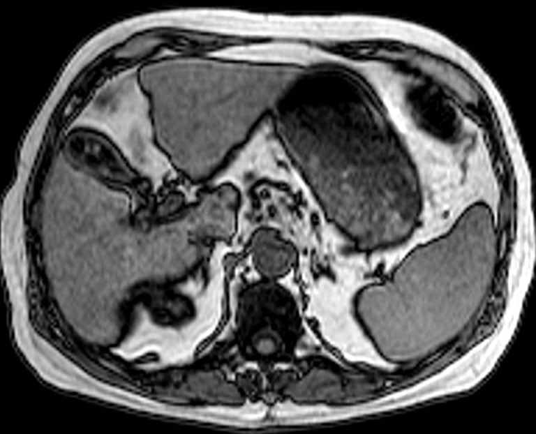
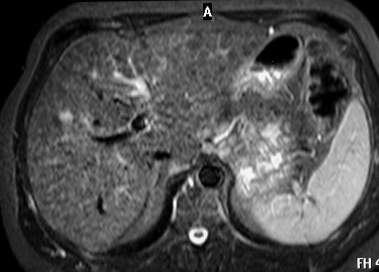
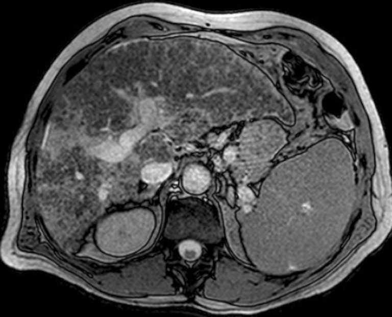
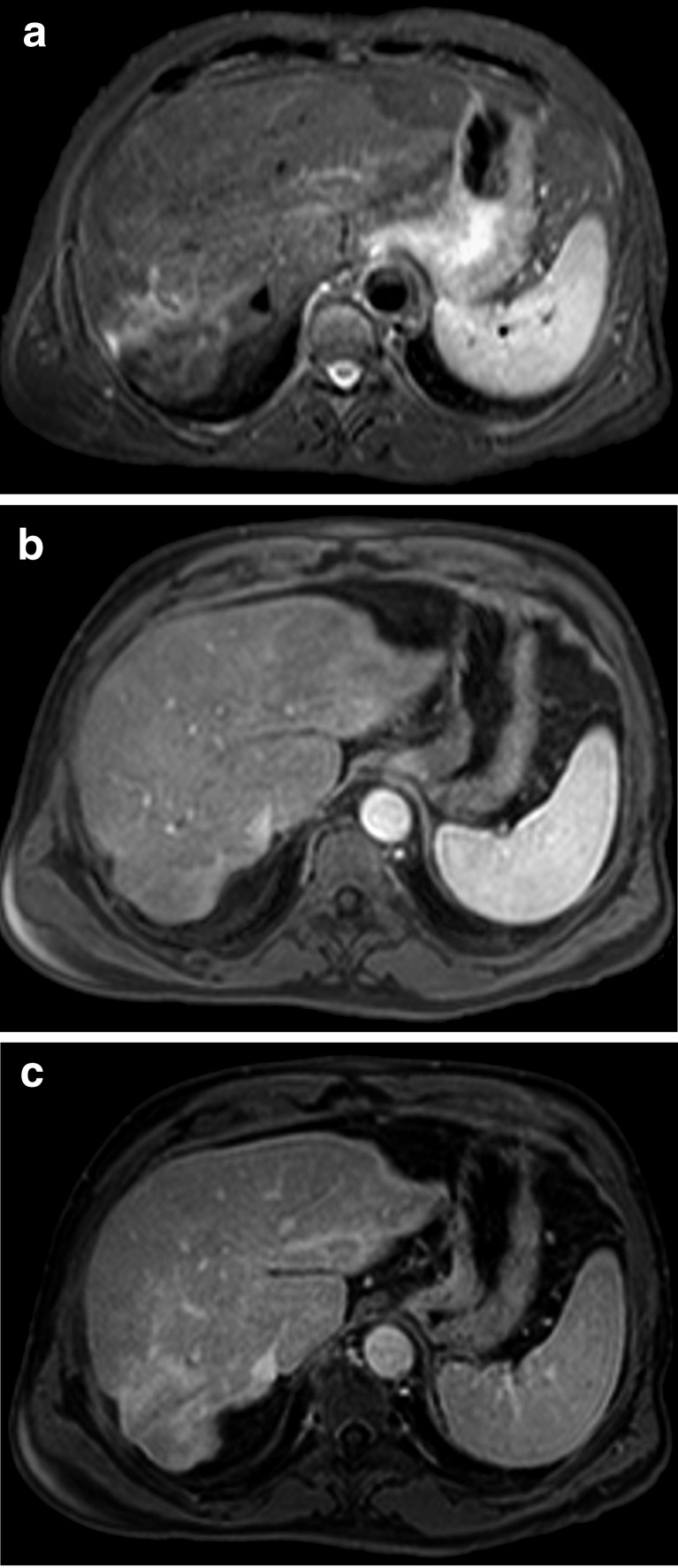
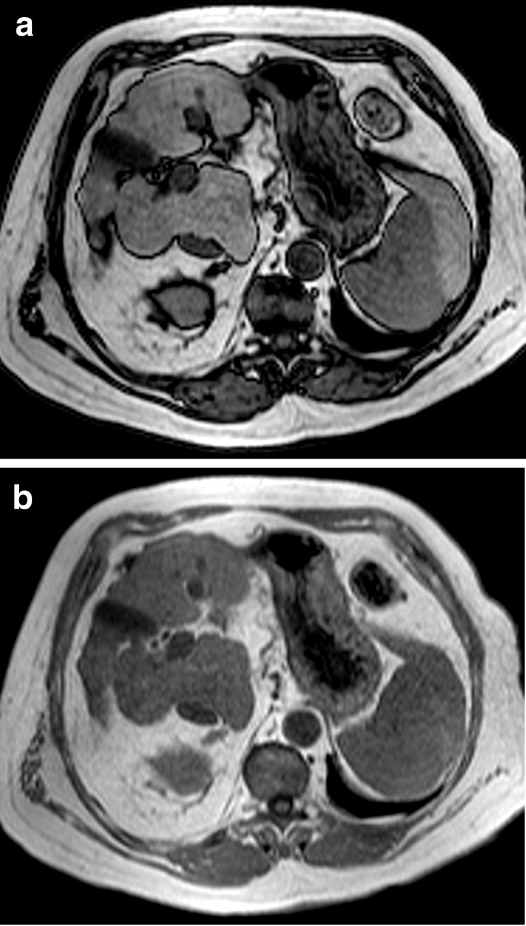
The typical histological features of chronic hepatitis and cirrhosis are variable degrees of hepatocellular necrosis and inflammation (activity or grade of disease), fibrosis (stage of disease), and associated fat and iron deposition. These features influence the liver's appearance and must be assessed separately by imaging biomarkers in order to be clinically useful. Hepatic morphologic alterations and features of portal hypertension identify most cases of established cirrhosis. Nowadays, research is focused on developing ways to improve detection of early and intermediate stages of fibrosis as well as hepatocyte dysfunction. Even more, most imaging-related measurements are subject to complex interactions and are influenced by different pathologic processes, such as fatty infiltration, edema, necrosis and iron accumulation. METHODS AND RESULTS: MR experience throughout the last 15 years at the Dr Peset University Hospital is reviewed. CONCLUSION: Nowadays, several biomarkers have been developed to grade the liver status in cirrhosis. This review will focus on these topics.

摘要

目的

慢性肝炎和肝硬化的典型组织学特征包括不同程度的肝细胞坏死和炎症(疾病活动度或分级)、纤维化(疾病分期)以及相关的脂肪和铁沉积。这些特征会影响肝脏的外观,为了具有临床实用性,必须通过成像生物标志物分别进行评估。肝脏形态学改变和门静脉高压特征可识别大多数已确诊的肝硬化病例。如今,研究集中在开发改善早期和中期纤维化以及肝细胞功能障碍检测的方法。此外,大多数与成像相关的测量都受到复杂相互作用的影响,并受到不同病理过程的影响,如脂肪浸润、水肿、坏死和铁蓄积。

方法与结果

回顾了佩塞特大学医院过去15年的磁共振成像经验。

结论

如今,已开发出多种生物标志物来对肝硬化的肝脏状态进行分级。本综述将聚焦于这些主题。

https://cdn.ncbi.nlm.nih.gov/pmc/blobs/913d/3288865/179b789fc48e/13244_2010_34_Fig17_HTML.jpg
https://cdn.ncbi.nlm.nih.gov/pmc/blobs/913d/3288865/0800a30f4ac7/13244_2010_34_Fig1_HTML.jpg
https://cdn.ncbi.nlm.nih.gov/pmc/blobs/913d/3288865/1c0626d707ea/13244_2010_34_Fig2_HTML.jpg
https://cdn.ncbi.nlm.nih.gov/pmc/blobs/913d/3288865/b7edd5fed6d2/13244_2010_34_Fig3_HTML.jpg
https://cdn.ncbi.nlm.nih.gov/pmc/blobs/913d/3288865/39c1a89874a2/13244_2010_34_Fig4_HTML.jpg
https://cdn.ncbi.nlm.nih.gov/pmc/blobs/913d/3288865/1204a061f921/13244_2010_34_Fig5_HTML.jpg
https://cdn.ncbi.nlm.nih.gov/pmc/blobs/913d/3288865/ed70b4f62445/13244_2010_34_Fig6_HTML.jpg
https://cdn.ncbi.nlm.nih.gov/pmc/blobs/913d/3288865/3154ce8ac246/13244_2010_34_Fig7_HTML.jpg
https://cdn.ncbi.nlm.nih.gov/pmc/blobs/913d/3288865/bf3f67608307/13244_2010_34_Fig8_HTML.jpg
https://cdn.ncbi.nlm.nih.gov/pmc/blobs/913d/3288865/c7ca5e49d056/13244_2010_34_Fig9_HTML.jpg
https://cdn.ncbi.nlm.nih.gov/pmc/blobs/913d/3288865/c176d2ef7a72/13244_2010_34_Fig10_HTML.jpg
https://cdn.ncbi.nlm.nih.gov/pmc/blobs/913d/3288865/5f906c2b010c/13244_2010_34_Fig11_HTML.jpg
https://cdn.ncbi.nlm.nih.gov/pmc/blobs/913d/3288865/98a63f5f53f0/13244_2010_34_Fig12_HTML.jpg
https://cdn.ncbi.nlm.nih.gov/pmc/blobs/913d/3288865/c94e2474fee3/13244_2010_34_Fig13_HTML.jpg
https://cdn.ncbi.nlm.nih.gov/pmc/blobs/913d/3288865/e640daddc3f2/13244_2010_34_Fig14_HTML.jpg
https://cdn.ncbi.nlm.nih.gov/pmc/blobs/913d/3288865/2115d7422b0e/13244_2010_34_Fig15_HTML.jpg
https://cdn.ncbi.nlm.nih.gov/pmc/blobs/913d/3288865/4f12d3eeb5a1/13244_2010_34_Fig16_HTML.jpg
https://cdn.ncbi.nlm.nih.gov/pmc/blobs/913d/3288865/179b789fc48e/13244_2010_34_Fig17_HTML.jpg

相似文献

1
MR imaging in liver cirrhosis: classical and new approaches.肝硬化的磁共振成像:传统方法与新方法
Insights Imaging. 2010 Sep;1(4):233-244. doi: 10.1007/s13244-010-0034-7. Epub 2010 Aug 5.
2
Ultrasound Shear Wave Elastography for Liver Disease. A Critical Appraisal of the Many Actors on the Stage.用于肝脏疾病的超声剪切波弹性成像:对该领域众多参与者的批判性评估
Ultraschall Med. 2016 Feb;37(1):1-5. doi: 10.1055/s-0035-1567037. Epub 2016 Feb 12.
3
Cirrhosis: MR imaging features.肝硬化:磁共振成像特征。
Magn Reson Imaging Clin N Am. 2002 Feb;10(1):75-92, vi. doi: 10.1016/s1064-9689(03)00050-3.
4
Imaging diagnosis of cirrhosis and chronic hepatitis.肝硬化和慢性肝炎的影像学诊断
Intervirology. 2004;47(3-5):134-43. doi: 10.1159/000078465.
5
Fibrosis and other histological features in chronic hepatitis C virus infection: a statistical model.丙型肝炎病毒慢性感染中的纤维化及其他组织学特征:一种统计模型
J Clin Pathol. 1996 Jun;49(6):465-9. doi: 10.1136/jcp.49.6.465.
6
Ductular reaction in hereditary hemochromatosis: the link between hepatocyte senescence and fibrosis progression.遗传性血色素沉着症中的小管反应:肝细胞衰老与纤维化进展之间的联系。
Hepatology. 2014 Mar;59(3):848-57. doi: 10.1002/hep.26706. Epub 2014 Jan 27.
7
Focus on histological abnormalities of intrahepatic vasculature in chronic viral hepatitis.关注慢性病毒性肝炎肝内血管组织学异常。
Liver Int. 2018 Oct;38(10):1770-1776. doi: 10.1111/liv.13718. Epub 2018 Mar 6.
8
Pathophysiology and a Rational Basis of Therapy.病理生理学与治疗的合理依据。
Dig Dis. 2015;33(4):508-14. doi: 10.1159/000374099. Epub 2015 Jul 6.
9
Imaging features of perivascular fatty infiltration of the liver: initial observations.肝脏血管周围脂肪浸润的影像学特征:初步观察
Radiology. 2005 Oct;237(1):159-69. doi: 10.1148/radiol.2371041580. Epub 2005 Aug 11.
10
Cirrhosis: spectrum of findings on unenhanced and dynamic gadolinium-enhanced MR imaging.肝硬化:未增强及动态钆增强磁共振成像的表现谱
Abdom Imaging. 2001 Nov-Dec;26(6):601-15. doi: 10.1007/s00261-001-0010-2.

引用本文的文献

1
Quantitative assessment of mucosal fibrosis and its correlation with disease severity and outcome in cats with chronic enteropathy.猫慢性肠病黏膜纤维化的定量评估及其与疾病严重程度和预后的相关性。
J Feline Med Surg. 2025 Jul;27(7):1098612X251338174. doi: 10.1177/1098612X251338174. Epub 2025 Jul 22.
2
MRI findings of confluent hepatic fibrosis caused by different etiologies.不同病因所致融合性肝纤维化的磁共振成像表现
BMC Gastroenterol. 2025 Jul 10;25(1):512. doi: 10.1186/s12876-025-04101-9.
3
Development of fully automated models for staging liver fibrosis using non-contrast MRI and artificial intelligence: a retrospective multicenter study.

本文引用的文献

1
MR spectroscopy of the liver: principles and clinical applications.肝脏磁共振波谱:原理与临床应用。
Radiographics. 2009 Oct;29(6):1653-64. doi: 10.1148/rg.296095520.
2
MR imaging of liver fibrosis: current state of the art.磁共振成像技术在肝纤维化中的应用:现状评估。
Radiographics. 2009 Oct;29(6):1615-35. doi: 10.1148/rg.296095512.
3
Advanced MRI methods for assessment of chronic liver disease.用于评估慢性肝病的先进磁共振成像方法。
使用非增强磁共振成像和人工智能开发用于肝纤维化分期的全自动模型:一项回顾性多中心研究
EClinicalMedicine. 2024 Oct 17;77:102881. doi: 10.1016/j.eclinm.2024.102881. eCollection 2024 Nov.
4
Quantitative evaluation of hepatic fibrosis by fibro Scan and Gd-EOB-DTPA-enhanced T1 mapping magnetic resonance imaging in chronic hepatitis B.肝纤维化的定量评估:磁共振成像 Gd-EOB-DTPA 增强 T1 mapping 与 FibroScan 在慢性乙型肝炎中的应用。
Abdom Radiol (NY). 2022 Feb;47(2):684-692. doi: 10.1007/s00261-021-03300-8. Epub 2021 Nov 25.
5
Magnetic Resonance imaging analysis of liver fibrosis and inflammation: overwhelming gray zones restrict clinical use.磁共振成像分析肝纤维化和炎症:压倒性的灰色地带限制了临床应用。
Abdom Radiol (NY). 2020 Nov;45(11):3557-3568. doi: 10.1007/s00261-020-02713-1. Epub 2020 Aug 28.
6
Transplantation of fetal liver tissue coated by ultra-purified alginate gel over liver improves hepatic function in the cirrhosis rat model.超纯藻酸盐凝胶包裹胎肝组织移植可改善肝硬化大鼠模型的肝功能。
Sci Rep. 2020 May 19;10(1):8231. doi: 10.1038/s41598-020-65069-y.
7
CEUS LI-RADS: a pictorial review.CEUS LI-RADS:图文综述。
Insights Imaging. 2020 Feb 4;11(1):9. doi: 10.1186/s13244-019-0819-2.
8
Magnetic Resonance Elastography of Liver: Current Update.肝脏磁共振弹性成像:最新进展
Top Magn Reson Imaging. 2018 Oct;27(5):319-333. doi: 10.1097/RMR.0000000000000177.
9
Putting it all together: established and emerging MRI techniques for detecting and measuring liver fibrosis.综合概述:用于检测和测量肝纤维化的成熟及新兴磁共振成像技术
Pediatr Radiol. 2018 Aug;48(9):1256-1272. doi: 10.1007/s00247-018-4083-2. Epub 2018 Aug 4.
AJR Am J Roentgenol. 2009 Jul;193(1):14-27. doi: 10.2214/AJR.09.2601.
4
Focal confluent fibrosis in cirrhotic liver: natural history studied with serial CT.肝硬化肝脏中的局灶性融合性纤维化:通过系列CT研究其自然史
AJR Am J Roentgenol. 2009 May;192(5):1341-7. doi: 10.2214/AJR.07.2782.
5
[Diagnosis and treatment of hepatocellular carcinoma].[肝细胞癌的诊断与治疗]
Med Clin (Barc). 2009 Feb 28;132(7):272-87. doi: 10.1016/j.medcli.2008.11.024. Epub 2009 Feb 14.
6
Evaluating the severity of nonalcoholic steatohepatitis with superparamagnetic iron oxide-enhanced magnetic resonance imaging.
J Magn Reson Imaging. 2008 Dec;28(6):1444-50. doi: 10.1002/jmri.21596.
7
Nonalcoholic fatty liver disease: rapid evaluation of liver fat content with in-phase and out-of-phase MR imaging.非酒精性脂肪性肝病:利用同相位和反相位磁共振成像快速评估肝脏脂肪含量
Radiology. 2009 Jan;250(1):130-6. doi: 10.1148/radiol.2501071934. Epub 2008 Nov 18.
8
Liver cirrhosis: intravoxel incoherent motion MR imaging--pilot study.肝硬化:体素内不相干运动磁共振成像——初步研究。
Radiology. 2008 Dec;249(3):891-9. doi: 10.1148/radiol.2493080080.
9
CT of nonneoplastic hepatic vascular and perfusion disorders.非肿瘤性肝血管及灌注异常的CT表现
Radiographics. 2008 Nov-Dec;28(7):1967-82. doi: 10.1148/rg.287085067.
10
Frequency and histopathologic basis of hepatic surface nodularity in patients with fulminant hepatic failure.暴发性肝衰竭患者肝表面结节的发生率及组织病理学基础
Radiology. 2008 Nov;249(2):518-23. doi: 10.1148/radiol.2492072168.