• 文献检索
  • 文档翻译
  • 深度研究
  • 学术资讯
  • Suppr Zotero 插件Zotero 插件
  • 邀请有礼
  • 套餐&价格
  • 历史记录
应用&插件
Suppr Zotero 插件Zotero 插件浏览器插件Mac 客户端Windows 客户端微信小程序
定价
高级版会员购买积分包购买API积分包
服务
文献检索文档翻译深度研究API 文档MCP 服务
关于我们
关于 Suppr公司介绍联系我们用户协议隐私条款
关注我们

Suppr 超能文献

核心技术专利:CN118964589B侵权必究
粤ICP备2023148730 号-1Suppr @ 2026

文献检索

告别复杂PubMed语法,用中文像聊天一样搜索,搜遍4000万医学文献。AI智能推荐,让科研检索更轻松。

立即免费搜索

文件翻译

保留排版,准确专业,支持PDF/Word/PPT等文件格式,支持 12+语言互译。

免费翻译文档

深度研究

AI帮你快速写综述,25分钟生成高质量综述,智能提取关键信息,辅助科研写作。

立即免费体验

使用非增强磁共振成像和人工智能开发用于肝纤维化分期的全自动模型:一项回顾性多中心研究

Development of fully automated models for staging liver fibrosis using non-contrast MRI and artificial intelligence: a retrospective multicenter study.

作者信息

Li Chunli, Wang Yuan, Bai Ruobing, Zhao Zhiyong, Li Wenjuan, Zhang Qianqian, Zhang Chaoya, Yang Wei, Liu Qi, Su Na, Lu Yueyue, Yin Xiaoli, Wang Fan, Gu Chengli, Yang Aoran, Luo Baihe, Zhou Minghui, Shen Liuhanxu, Pan Chen, Wang Zhiying, Wu Qijun, Yin Jiandong, Hou Yang, Shi Yu

机构信息

Department of Radiology, Shengjing Hospital of China Medical University, Shenyang, Liaoning, China.

Department of Radiology, Cancer Hospital of China Medical University, Liaoning Cancer Hospital & Institute, Shenyang, Liaoning, China.

出版信息

EClinicalMedicine. 2024 Oct 17;77:102881. doi: 10.1016/j.eclinm.2024.102881. eCollection 2024 Nov.

DOI:10.1016/j.eclinm.2024.102881
PMID:39498462
原文链接:https://pmc.ncbi.nlm.nih.gov/articles/PMC11532432/
Abstract

BACKGROUND

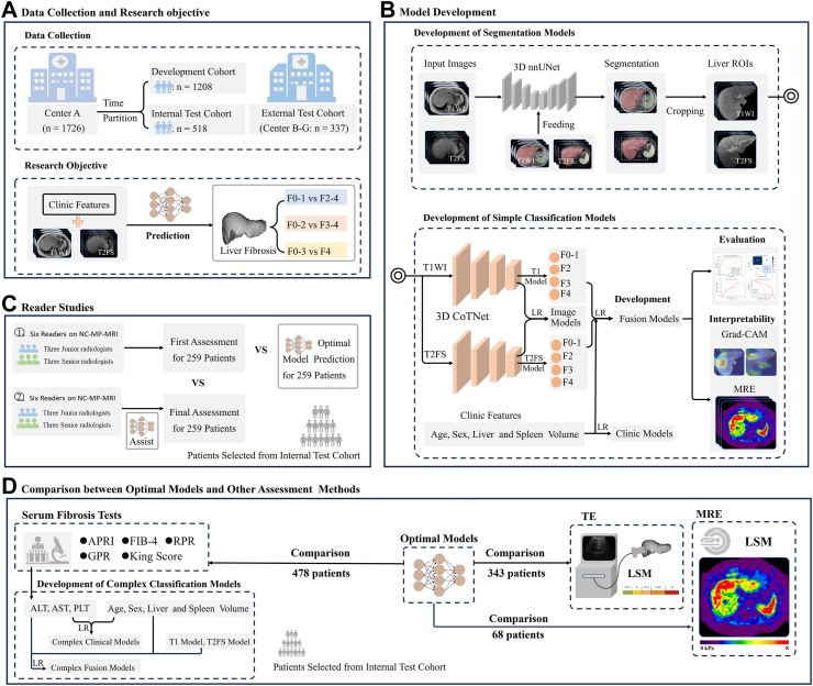
Accurate staging of liver fibrosis (LF) is essential for clinical management in chronic liver disease. While non-contrast MRI (NC-MRI) yields valuable information for liver assessment, its effectiveness in predicting LF remains underexplored. This study aimed to develop and validate artificial intelligence (AI)-powered models utilizing NC-MRI for staging LF.

METHODS

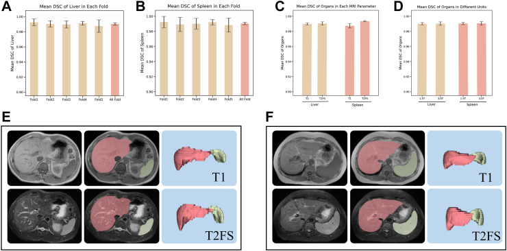
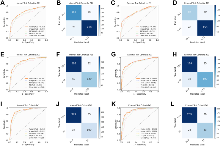
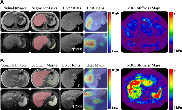
A total of 1726 patients from Shengjing Hospital of China Medical University, registered between October 2003 and October 2022, were retrospectively collected, and divided into development (n = 1208) and internal test (n = 518) cohorts. An external test cohort consisting of 337 individuals from six centers, registered between June 2015 and November 2022, were also included. All participants underwent NC-MRI (T1-weighted imaging, T1WI; and T2-fat-suppressed imaging, T2FS) and liver biopsies. Two classification models (CMs), named T1 and T2FS, were trained on respective image types using 3D contextual transformer networks and evaluated on both test cohorts. Additionally, three CMs-Clinic, Image, and Fusion-were developed using clinical features, T1 and T2FS scores, and their integration via logistic regression. Classification effectiveness of CMs was assessed using the area under the receiver operating characteristic curve (AUC). A comparison was conducted between the optimal models (OMs) with highest AUC and other methods (transient elastography, five serum biomarkers, and six radiologists).

FINDINGS

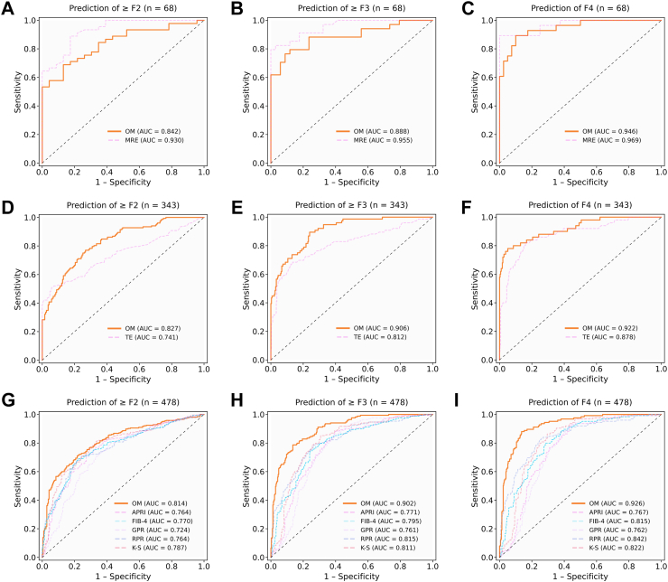
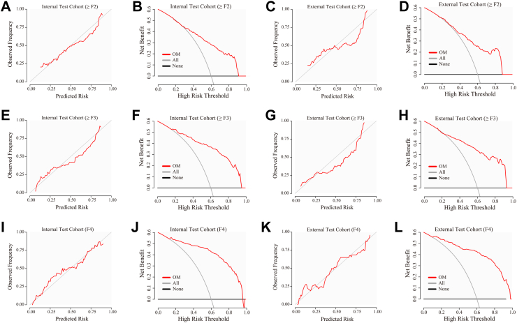
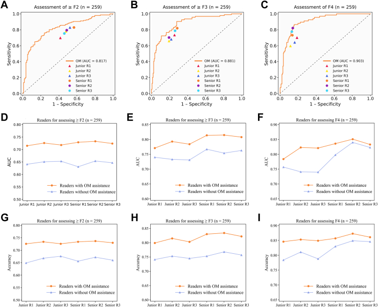
Fusion models (i.e., OM) yielded the highest AUC among the CMs, achieving AUCs of 0.810 for significant fibrosis, 0.881 for advanced fibrosis, and 0.918 for cirrhosis in the internal test cohort, and 0.808, 0.868, and 0.925, respectively, in the external test cohort. The OMs demonstrated superior performance in AUC, significantly surpassing transient elastography (only for staging ≥ F2 and ≥ F3 grades), serum biomarkers, and three junior radiologists for staging LF. Radiologists, with the aid of the OMs, can achieve a higher AUC in LF assessment.

INTERPRETATION

AI-powered models utilizing NC-MRI, including T1WI and T2FS, accurately stage LF.

FUNDING

National Natural Science Foundation of China (No. 82071885); General Program of the Liaoning Provincial Department of Education (LJKMZ20221160); Liaoning Province Science and Technology Joint Plan (2023JH2/101700127); the Leading Young Talent Program of Xingliao Yingcai in Liaoning Province (XLYC2203037).

摘要

背景

肝纤维化(LF)的准确分期对于慢性肝病的临床管理至关重要。虽然非增强MRI(NC-MRI)可为肝脏评估提供有价值的信息,但其在预测LF方面的有效性仍未得到充分探索。本研究旨在开发并验证利用NC-MRI进行LF分期的人工智能(AI)模型。

方法

回顾性收集了2003年10月至2022年10月在中国医科大学附属盛京医院登记的1726例患者,并将其分为开发队列(n = 1208)和内部测试队列(n = 518)。还纳入了一个由来自六个中心的337名个体组成的外部测试队列,这些个体于2015年6月至2022年11月登记。所有参与者均接受了NC-MRI(T1加权成像,T1WI;以及T2脂肪抑制成像,T2FS)和肝脏活检。使用3D上下文变压器网络在各自的图像类型上训练了两个分类模型(CMs),分别命名为T1和T2FS,并在两个测试队列上进行评估。此外,使用临床特征、T1和T2FS评分以及通过逻辑回归对它们进行整合,开发了三个CMs——临床、图像和融合模型。使用受试者操作特征曲线(AUC)下的面积评估CMs的分类有效性。在具有最高AUC的最佳模型(OMs)与其他方法(瞬时弹性成像、五种血清生物标志物和六名放射科医生)之间进行了比较。

结果

融合模型(即OMs)在CMs中产生了最高的AUC,在内部测试队列中,显著纤维化的AUC为0.810,晚期纤维化的AUC为0.881,肝硬化的AUC为0.918,在外部测试队列中分别为0.808、0.868和0.925。OMs在AUC方面表现出卓越的性能,在LF分期方面显著超过瞬时弹性成像(仅用于分期≥F2和≥F3级)、血清生物标志物以及三名初级放射科医生。放射科医生借助OMs在LF评估中可以获得更高的AUC。

解读

利用包括T1WI和T2FS在内的NC-MRI的AI模型能够准确地对LF进行分期。

资助

国家自然科学基金(编号82071885);辽宁省教育厅一般项目(LJKMZ20221160);辽宁省科技联合计划(2023JH2/101700127);辽宁省兴辽英才青年拔尖人才计划(XLYC2203037)。

https://cdn.ncbi.nlm.nih.gov/pmc/blobs/0cfe/11532432/fbd362cef2ad/gr7.jpg
https://cdn.ncbi.nlm.nih.gov/pmc/blobs/0cfe/11532432/7bc43444d3de/gr1.jpg
https://cdn.ncbi.nlm.nih.gov/pmc/blobs/0cfe/11532432/b8d955d8a977/gr2.jpg
https://cdn.ncbi.nlm.nih.gov/pmc/blobs/0cfe/11532432/9fa329d63db0/gr3.jpg
https://cdn.ncbi.nlm.nih.gov/pmc/blobs/0cfe/11532432/b709d6c11f2c/gr4.jpg
https://cdn.ncbi.nlm.nih.gov/pmc/blobs/0cfe/11532432/4ef1060bec2d/gr5.jpg
https://cdn.ncbi.nlm.nih.gov/pmc/blobs/0cfe/11532432/37feee75a2cb/gr6.jpg
https://cdn.ncbi.nlm.nih.gov/pmc/blobs/0cfe/11532432/fbd362cef2ad/gr7.jpg

相似文献

1
Development of fully automated models for staging liver fibrosis using non-contrast MRI and artificial intelligence: a retrospective multicenter study.使用非增强磁共振成像和人工智能开发用于肝纤维化分期的全自动模型:一项回顾性多中心研究
EClinicalMedicine. 2024 Oct 17;77:102881. doi: 10.1016/j.eclinm.2024.102881. eCollection 2024 Nov.
2
Comparing and combining MRE, T1ρ, SWI, IVIM, and DCE-MRI for the staging of liver fibrosis in rabbits: Assessment of a predictive model based on multiparametric MRI.比较和结合 MRE、T1ρ、SWI、IVIM 和 DCE-MRI 对兔肝纤维化分期:基于多参数 MRI 的预测模型评估。
Magn Reson Med. 2022 May;87(5):2424-2435. doi: 10.1002/mrm.29126. Epub 2021 Dec 21.
3
Multiparametric MRI-based whole-liver radiomics for predicting early-stage liver fibrosis in rabbits.基于多参数 MRI 的全肝放射组学预测兔早期肝纤维化
Br J Radiol. 2024 May 7;97(1157):964-970. doi: 10.1093/bjr/tqae063.
4
Diffusion-weighted MRI-based Virtual Elastography for the Assessment of Liver Fibrosis.基于扩散加权磁共振成像的虚拟弹性成像用于评估肝纤维化。
Radiology. 2020 Apr;295(1):127-135. doi: 10.1148/radiol.2020191498. Epub 2020 Feb 11.
5
Fully automated hybrid approach on conventional MRI for triaging clinically significant liver fibrosis: A multi-center cohort study.基于常规 MRI 的全自动混合方法对临床显著肝纤维化进行分诊:一项多中心队列研究。
J Med Virol. 2024 Aug;96(8):e29882. doi: 10.1002/jmv.29882.
6
Early prediction of treatment response to neoadjuvant chemotherapy based on longitudinal ultrasound images of HER2-positive breast cancer patients by Siamese multi-task network: A multicentre, retrospective cohort study.基于连体多任务网络的HER2阳性乳腺癌患者纵向超声图像对新辅助化疗治疗反应的早期预测:一项多中心回顾性队列研究
EClinicalMedicine. 2022 Jul 30;52:101562. doi: 10.1016/j.eclinm.2022.101562. eCollection 2022 Oct.
7
Prospective comparison of magnetic resonance imaging to transient elastography and serum markers for liver fibrosis detection.磁共振成像与瞬时弹性成像及血清标志物用于肝纤维化检测的前瞻性比较
Liver Int. 2016 May;36(5):659-66. doi: 10.1111/liv.13058. Epub 2016 Feb 7.
8
Highly sensitive detection platform-based diagnosis of oesophageal squamous cell carcinoma in China: a multicentre, case-control, diagnostic study.基于高灵敏度检测平台的中国食管鳞状细胞癌诊断:一项多中心、病例对照诊断研究。
Lancet Digit Health. 2024 Oct;6(10):e705-e717. doi: 10.1016/S2589-7500(24)00153-5.
9
Diagnostic accuracy and clinical impact of MRI-based technologies for patients with non-alcoholic fatty liver disease: systematic review and economic evaluation.基于 MRI 的技术在非酒精性脂肪性肝病患者中的诊断准确性和临床影响:系统评价和经济评估。
Health Technol Assess. 2023 Jul;27(10):1-115. doi: 10.3310/KGJU3398.
10
Deep learning-enabled fully automated pipeline system for segmentation and classification of single-mass breast lesions using contrast-enhanced mammography: a prospective, multicentre study.使用对比增强乳腺X线摄影术的深度学习驱动的单灶性乳腺病变分割与分类全自动管道系统:一项前瞻性多中心研究
EClinicalMedicine. 2023 Mar 17;58:101913. doi: 10.1016/j.eclinm.2023.101913. eCollection 2023 Apr.

引用本文的文献

1
Nutrients as epigenetic modulators in metabolic dysfunction-associated steatotic liver disease.营养素作为代谢功能障碍相关脂肪性肝病中的表观遗传调节剂。
World J Hepatol. 2025 Aug 27;17(8):108182. doi: 10.4254/wjh.v17.i8.108182.
2
Aortic atherosclerosis evaluation using deep learning based on non-contrast CT: A retrospective multi-center study.基于非增强CT的深度学习用于主动脉粥样硬化评估:一项回顾性多中心研究。
iScience. 2025 Jul 12;28(8):113100. doi: 10.1016/j.isci.2025.113100. eCollection 2025 Aug 15.
3
A large language model based pipeline for extracting information from patient complaint and anamnesis in clinical notes for severity assessment.

本文引用的文献

1
Machine Learning-Based Models for Advanced Fibrosis and Cirrhosis Diagnosis in Chronic Hepatitis B Patients With Hepatic Steatosis.基于机器学习的模型在脂肪性肝炎慢性乙型肝炎患者肝纤维化和肝硬化诊断中的应用。
Clin Gastroenterol Hepatol. 2024 Nov;22(11):2250-2260.e12. doi: 10.1016/j.cgh.2024.06.014. Epub 2024 Jun 19.
2
Accuracy of blood-based biomarkers for staging liver fibrosis in chronic liver disease: A systematic review supporting the AASLD Practice Guideline.用于慢性肝病肝纤维化分期的血液生物标志物准确性:支持美国肝病研究学会实践指南的系统评价
Hepatology. 2025 Jan 1;81(1):358-379. doi: 10.1097/HEP.0000000000000842. Epub 2024 Mar 15.
3
一种基于大语言模型的管道,用于从临床记录中的患者主诉和病史中提取信息以进行严重程度评估。
Sci Rep. 2025 Jul 14;15(1):25345. doi: 10.1038/s41598-025-07649-4.
4
[Fatigue in the general population: results of the "German Health Update 2023" study].[普通人群中的疲劳:“2023年德国健康更新”研究结果]
Bundesgesundheitsblatt Gesundheitsforschung Gesundheitsschutz. 2024 Nov;67(11):1208-1221. doi: 10.1007/s00103-024-03950-1. Epub 2024 Sep 26.
Noninvasive diagnosis of liver cirrhosis: qualitative and quantitative imaging biomarkers.
肝硬化的非侵入性诊断:定性和定量成像生物标志物
Abdom Radiol (NY). 2024 Jun;49(6):2098-2115. doi: 10.1007/s00261-024-04225-8. Epub 2024 Feb 19.
4
MASLD: a systemic metabolic disorder with cardiovascular and malignant complications.代谢相关脂肪性肝病:一种伴有心血管和恶性并发症的全身性代谢紊乱疾病。
Gut. 2024 Mar 7;73(4):691-702. doi: 10.1136/gutjnl-2023-330595.
5
Automated liver segmental volume ratio quantification on non-contrast T1-Vibe Dixon liver MRI using deep learning.基于深度学习的非对比 T1-Vibe Dixon 肝脏 MRI 自动肝段容积比定量
Eur J Radiol. 2023 Oct;167:111047. doi: 10.1016/j.ejrad.2023.111047. Epub 2023 Aug 14.
6
Global epidemiology of cirrhosis - aetiology, trends and predictions.全球肝硬化的流行病学:病因、趋势和预测。
Nat Rev Gastroenterol Hepatol. 2023 Jun;20(6):388-398. doi: 10.1038/s41575-023-00759-2. Epub 2023 Mar 28.
7
Diagnosis and management of autoimmune hepatitis.自身免疫性肝炎的诊断与管理
BMJ. 2023 Feb 6;380:e070201. doi: 10.1136/bmj-2022-070201.
8
Fully Automated and Explainable Liver Segmental Volume Ratio and Spleen Segmentation at CT for Diagnosing Cirrhosis.CT 上用于诊断肝硬化的全自动且可解释的肝脏节段体积比及脾脏分割
Radiol Artif Intell. 2022 Aug 24;4(5):e210268. doi: 10.1148/ryai.210268. eCollection 2022 Sep.
9
Hepatocellular Carcinoma Incidence in Alcohol-Associated Cirrhosis: Systematic Review and Meta-analysis.酒精性肝硬化相关肝细胞癌的发病率:系统评价和荟萃分析。
Clin Gastroenterol Hepatol. 2023 May;21(5):1169-1177. doi: 10.1016/j.cgh.2022.06.032. Epub 2022 Aug 5.
10
Contextual Transformer Networks for Visual Recognition.用于视觉识别的上下文Transformer网络
IEEE Trans Pattern Anal Mach Intell. 2023 Feb;45(2):1489-1500. doi: 10.1109/TPAMI.2022.3164083. Epub 2023 Jan 6.