• 文献检索
  • 文档翻译
  • 深度研究
  • 学术资讯
  • Suppr Zotero 插件Zotero 插件
  • 邀请有礼
  • 套餐&价格
  • 历史记录
应用&插件
Suppr Zotero 插件Zotero 插件浏览器插件Mac 客户端Windows 客户端微信小程序
定价
高级版会员购买积分包购买API积分包
服务
文献检索文档翻译深度研究API 文档MCP 服务
关于我们
关于 Suppr公司介绍联系我们用户协议隐私条款
关注我们

Suppr 超能文献

核心技术专利:CN118964589B侵权必究
粤ICP备2023148730 号-1Suppr @ 2026

文献检索

告别复杂PubMed语法,用中文像聊天一样搜索,搜遍4000万医学文献。AI智能推荐,让科研检索更轻松。

立即免费搜索

文件翻译

保留排版,准确专业,支持PDF/Word/PPT等文件格式,支持 12+语言互译。

免费翻译文档

深度研究

AI帮你快速写综述,25分钟生成高质量综述,智能提取关键信息,辅助科研写作。

立即免费体验

脊柱CT成像的技术层面

Technical aspects of CT imaging of the spine.

作者信息

Tins Bernhard

机构信息

Department of Radiology, RJAH Orthopaedic Hospital, Oswestry, Shropshire SY 107 AG UK.

出版信息

Insights Imaging. 2010 Nov;1(5-6):349-359. doi: 10.1007/s13244-010-0047-2. Epub 2010 Oct 21.

DOI:10.1007/s13244-010-0047-2
PMID:22347928
原文链接:https://pmc.ncbi.nlm.nih.gov/articles/PMC3259341/
Abstract

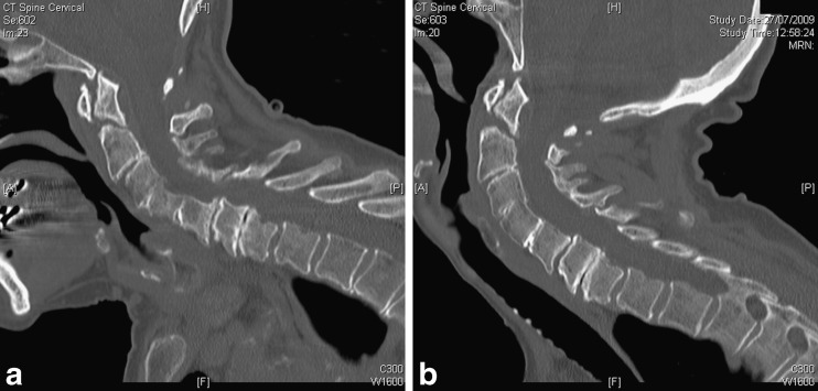
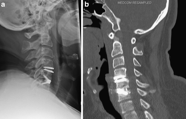
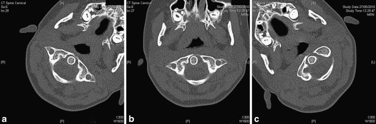
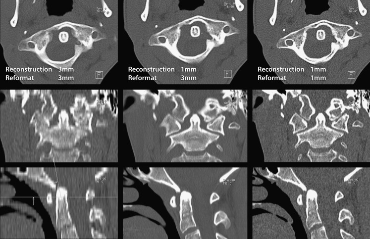
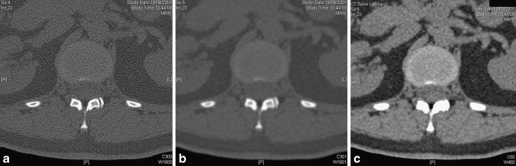
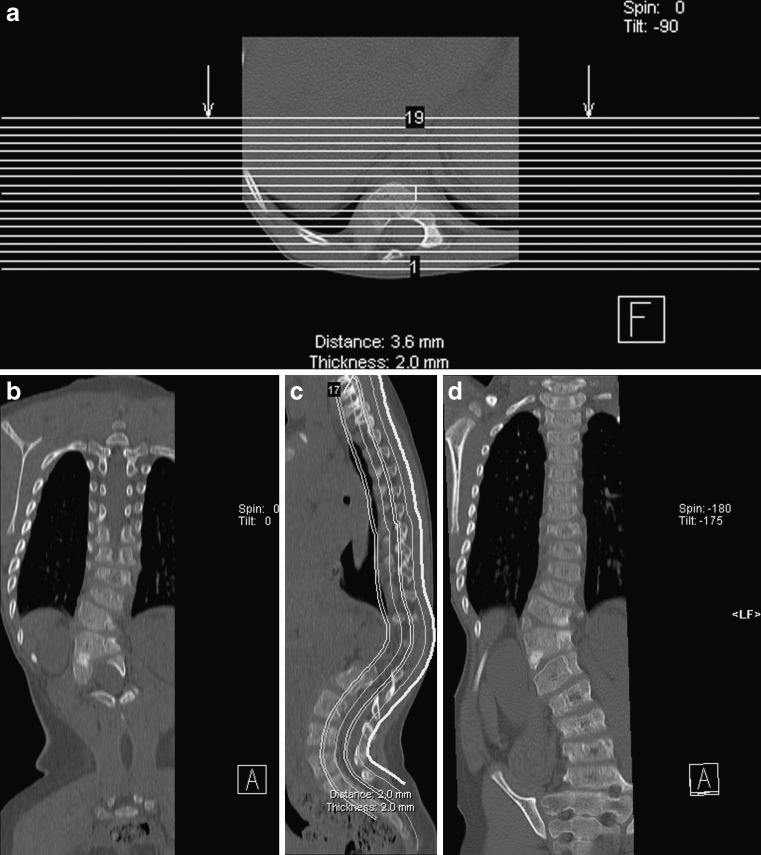
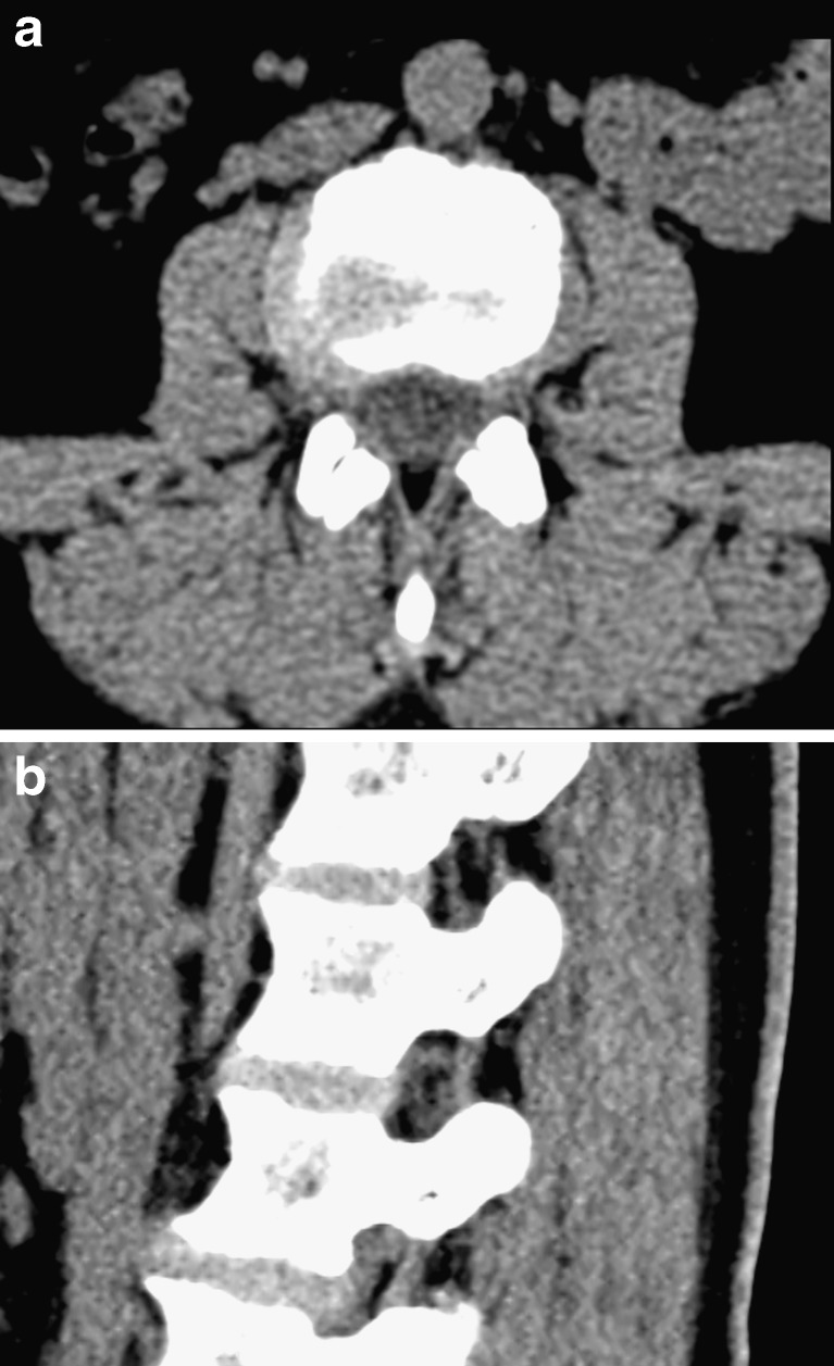
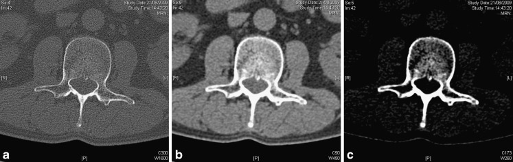
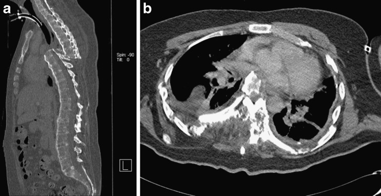
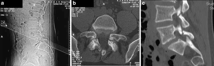
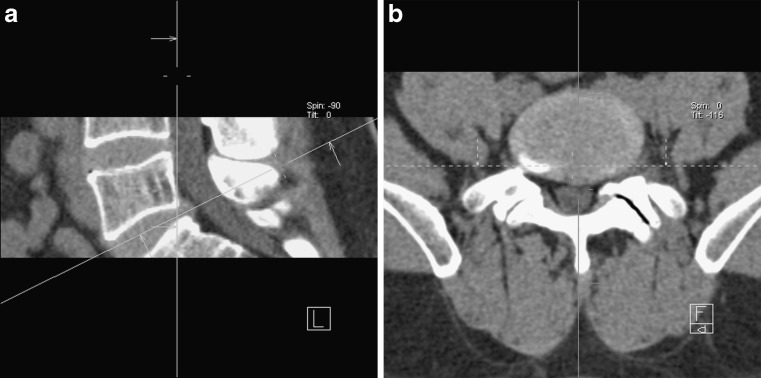
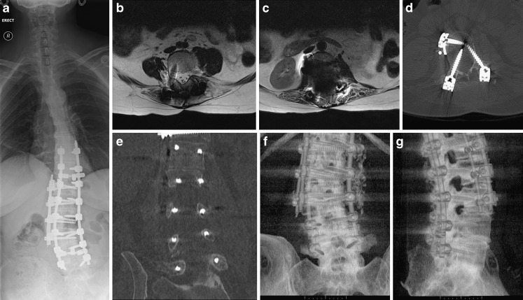
This review article discusses technical aspects of computed tomography (CT) imaging of the spine. Patient positioning, and its influence on image quality and movement artefact, is discussed. Particular emphasis is placed on the choice of scan parameters and their relation to image quality and radiation burden to the patient. Strategies to reduce radiation burden and artefact from metal implants are outlined. Data acquisition, processing, image display and steps to reduce artefact are reviewed. CT imaging of the spine is put into context with other imaging modalities for specific clinical indications or problems. This review aims to review underlying principles for image acquisition and to provide a rough guide for clinical problems without being prescriptive. Individual practice will always vary and reflect differences in local experience, technical provisions and clinical requirements.

摘要

这篇综述文章讨论了脊柱计算机断层扫描(CT)成像的技术方面。探讨了患者体位及其对图像质量和运动伪影的影响。特别强调了扫描参数的选择及其与图像质量和患者辐射负担的关系。概述了减少金属植入物辐射负担和伪影的策略。回顾了数据采集、处理、图像显示以及减少伪影的步骤。针对特定临床指征或问题,将脊柱CT成像与其他成像方式进行了对比。本综述旨在回顾图像采集的基本原则,并为临床问题提供一个大致的指导,而非给出具体的规定。个人实践总会有所不同,反映出当地经验、技术条件和临床需求的差异。

https://cdn.ncbi.nlm.nih.gov/pmc/blobs/ea14/3288870/96602f87fdaa/13244_2010_47_Fig12_HTML.jpg
https://cdn.ncbi.nlm.nih.gov/pmc/blobs/ea14/3288870/397a725dd312/13244_2010_47_Fig1_HTML.jpg
https://cdn.ncbi.nlm.nih.gov/pmc/blobs/ea14/3288870/37da339bc4d6/13244_2010_47_Fig2_HTML.jpg
https://cdn.ncbi.nlm.nih.gov/pmc/blobs/ea14/3288870/0e90f23c5a48/13244_2010_47_Fig3_HTML.jpg
https://cdn.ncbi.nlm.nih.gov/pmc/blobs/ea14/3288870/48f0ab3b9b25/13244_2010_47_Fig4_HTML.jpg
https://cdn.ncbi.nlm.nih.gov/pmc/blobs/ea14/3288870/b7e3d5f51f9a/13244_2010_47_Fig5_HTML.jpg
https://cdn.ncbi.nlm.nih.gov/pmc/blobs/ea14/3288870/ad2d456302e5/13244_2010_47_Fig6_HTML.jpg
https://cdn.ncbi.nlm.nih.gov/pmc/blobs/ea14/3288870/a3b1de36d4f8/13244_2010_47_Fig7_HTML.jpg
https://cdn.ncbi.nlm.nih.gov/pmc/blobs/ea14/3288870/6e50c7ad4830/13244_2010_47_Fig8_HTML.jpg
https://cdn.ncbi.nlm.nih.gov/pmc/blobs/ea14/3288870/b1e10a0be0e3/13244_2010_47_Fig9_HTML.jpg
https://cdn.ncbi.nlm.nih.gov/pmc/blobs/ea14/3288870/26a8610d1fa0/13244_2010_47_Fig10_HTML.jpg
https://cdn.ncbi.nlm.nih.gov/pmc/blobs/ea14/3288870/818aa5ad1787/13244_2010_47_Fig11_HTML.jpg
https://cdn.ncbi.nlm.nih.gov/pmc/blobs/ea14/3288870/96602f87fdaa/13244_2010_47_Fig12_HTML.jpg

相似文献

1
Technical aspects of CT imaging of the spine.脊柱CT成像的技术层面
Insights Imaging. 2010 Nov;1(5-6):349-359. doi: 10.1007/s13244-010-0047-2. Epub 2010 Oct 21.
2
CT and MRI Techniques for Imaging Around Orthopedic Hardware.用于骨科植入物周围成像的CT和MRI技术
Rofo. 2018 Jan;190(1):31-41. doi: 10.1055/s-0043-118127. Epub 2017 Sep 21.
3
Post-mortem computed tomography: Technical principles and recommended parameter settings for high-resolution imaging.尸检计算机断层扫描:高分辨率成像的技术原理及推荐参数设置
Med Sci Law. 2018 Jan;58(1):70-82. doi: 10.1177/0025802417747167. Epub 2018 Jan 8.
4
Metallic artefact reduction with monoenergetic dual-energy CT: systematic ex vivo evaluation of posterior spinal fusion implants from various vendors and different spine levels.单能量双能 CT 降低金属伪影:来自不同供应商和不同脊柱水平的后路脊柱融合植入物的系统离体评估。
Eur Radiol. 2012 Nov;22(11):2357-64. doi: 10.1007/s00330-012-2501-7. Epub 2012 May 30.
5
Technical Pitfalls and Limitations of SPECT/CT.单光子发射计算机断层扫描/计算机断层扫描(SPECT/CT)的技术陷阱与局限性
Semin Nucl Med. 2015 Nov;45(6):530-40. doi: 10.1053/j.semnuclmed.2015.06.002.
6
Radiation dose-reduction strategies in thoracic CT.胸部CT中的辐射剂量降低策略。
Clin Radiol. 2017 May;72(5):407-420. doi: 10.1016/j.crad.2016.11.021. Epub 2017 Jan 27.
7
Integration of FDG-PET/CT into external beam radiation therapy planning: technical aspects and recommendations on methodological approaches.将氟代脱氧葡萄糖正电子发射断层显像/计算机断层扫描(FDG-PET/CT)整合到外照射放射治疗计划中:技术方面及方法学建议
Nuklearmedizin. 2012;51(4):140-53. doi: 10.3413/Nukmed-0455-11-12. Epub 2012 Apr 3.
8
Basics of iterative reconstruction methods in computed tomography: A vendor-independent overview.计算机断层扫描中迭代重建方法的基础:与供应商无关的概述。
Eur J Radiol. 2018 Dec;109:147-154. doi: 10.1016/j.ejrad.2018.10.025. Epub 2018 Oct 26.
9
Dosimetric impact of a CT metal artefact suppression algorithm for proton, electron and photon therapies.一种用于质子、电子和光子治疗的CT金属伪影抑制算法的剂量学影响。
Phys Med Biol. 2006 Oct 21;51(20):5183-97. doi: 10.1088/0031-9155/51/20/007. Epub 2006 Sep 27.
10
Deep learning-based metal artefact reduction in PET/CT imaging.基于深度学习的 PET/CT 成像中的金属伪影减少。
Eur Radiol. 2021 Aug;31(8):6384-6396. doi: 10.1007/s00330-021-07709-z. Epub 2021 Feb 10.

引用本文的文献

1
Diagnostic performance of lumbar spine CT using deep learning denoising to evaluate disc herniation and spinal stenosis.使用深度学习去噪的腰椎CT对椎间盘突出和椎管狭窄的诊断性能
Eur Radiol. 2025 Jun 7. doi: 10.1007/s00330-025-11742-7.
2
Spinal navigation with AI-driven 3D-reconstruction of fluoroscopy images: an ex-vivo feasibility study.基于人工智能的透视图像三维重建的脊柱导航:一项离体可行性研究。
BMC Musculoskelet Disord. 2024 Nov 19;25(1):925. doi: 10.1186/s12891-024-08052-2.
3
Towards a standardized reporting of the impact of magnetic resonance imaging on the decision-making of thoracolumbar fractures without neurological deficit: Conceptual framework and proposed methodology.

本文引用的文献

1
Radiation Dose Optimization in CT Planning of Corrective Scoliosis Surgery. A Phantom Study.矫正脊柱侧弯手术CT规划中的辐射剂量优化:一项模体研究
Neuroradiol J. 2008 Jun 3;21(3):374-82. doi: 10.1177/197140090802100313.
2
Quantitative computed tomography.定量计算机断层扫描
Radiol Clin North Am. 2010 May;48(3):589-600. doi: 10.1016/j.rcl.2010.03.001.
3
How effective is effective dose as a predictor of radiation risk?有效剂量作为辐射风险预测因子的有效性如何?
迈向标准化报告磁共振成像对无神经功能缺损的胸腰椎骨折决策影响的研究:概念框架与拟议方法
Brain Spine. 2024 Mar 21;4:102787. doi: 10.1016/j.bas.2024.102787. eCollection 2024.
4
Radiologic reporting of MRI-proven thoracolumbar epidural metastases on body CT: 12-Year single-institution experience.体部 CT 诊断胸腰椎硬膜外转移瘤的放射学报告:12 年单机构经验。
Clin Imaging. 2023 Oct;102:19-25. doi: 10.1016/j.clinimag.2023.06.025. Epub 2023 Jul 7.
5
Photon-Counting Computed Tomography (PC-CT) of the spine: impact on diagnostic confidence and radiation dose.光子计数 CT (PC-CT)在脊柱中的应用:对诊断信心和辐射剂量的影响。
Eur Radiol. 2023 Aug;33(8):5578-5586. doi: 10.1007/s00330-023-09511-5. Epub 2023 Mar 9.
6
Cervical spine injury: clinical and medico-legal overview.颈椎损伤:临床与医学法律综述。
Radiol Med. 2023 Jan;128(1):103-112. doi: 10.1007/s11547-022-01578-2. Epub 2023 Jan 31.
7
A Proposal for a Standardized Imaging Algorithm to Improve the Accuracy and Reliability for the Diagnosis of Thoracolumbar Posterior Ligamentous Complex Injury in Computed Tomography and Magnetic Resonance Imaging.一种标准化成像算法的建议,以提高计算机断层扫描和磁共振成像中胸腰椎后韧带复合体损伤诊断的准确性和可靠性。
Global Spine J. 2023 Apr;13(3):873-896. doi: 10.1177/21925682221129220. Epub 2022 Oct 12.
8
Evaluation of the contact surface between vertebral endplate and 3D printed patient-specific cage vs commercial cage.评估椎骨终板与 3D 打印个体化椎间融合器和市售椎间融合器之间的接触面。
Sci Rep. 2022 Jul 22;12(1):12505. doi: 10.1038/s41598-022-16895-9.
9
Diagnostic Value of Radioisotope Cisternography Using In-DTPA in a Patient with Rhinorrhea and Purulent Meningitis.放射性核素脑池造影术在鼻漏伴脓性脑膜炎患者中的诊断价值。
Medicina (Kaunas). 2022 May 26;58(6):714. doi: 10.3390/medicina58060714.
10
A morphometric analysis of thoracolumbar vertebrae in goat by computed tomography.山羊胸腰椎的计算机断层扫描形态计量学分析
Vet Anim Sci. 2022 Jan 17;15:100233. doi: 10.1016/j.vas.2022.100233. eCollection 2022 Mar.
AJR Am J Roentgenol. 2010 Apr;194(4):890-6. doi: 10.2214/AJR.09.4179.
4
Estimating effective dose for CT using dose-length product compared with using organ doses: consequences of adopting International Commission on Radiological Protection publication 103 or dual-energy scanning.使用剂量长度产品与使用器官剂量估算 CT 的有效剂量:采用国际放射防护委员会第 103 号出版物或双能扫描的后果。
AJR Am J Roentgenol. 2010 Apr;194(4):881-9. doi: 10.2214/AJR.09.3462.
5
Spine computed tomography doses and cancer induction.脊柱计算机断层扫描剂量与癌症诱导。
Spine (Phila Pa 1976). 2010 Feb 15;35(4):430-3. doi: 10.1097/BRS.0b013e3181cdde47.
6
Diagnostic CT radiation and cancer induction.诊断性CT辐射与癌症诱发。
Skeletal Radiol. 2010 May;39(5):421-4. doi: 10.1007/s00256-009-0819-2.
7
Quantitative computed tomography.定量计算机断层扫描
Eur J Radiol. 2009 Sep;71(3):415-24. doi: 10.1016/j.ejrad.2009.04.074. Epub 2009 Aug 13.
8
Radiation exposure from musculoskeletal computerized tomographic scans.肌肉骨骼计算机断层扫描的辐射暴露
J Bone Joint Surg Am. 2009 Aug;91(8):1882-9. doi: 10.2106/JBJS.H.01199.
9
Reliability of low-radiation dose CT in the assessment of screw placement after posterior scoliosis surgery, evaluated with a new grading system.采用一种新的分级系统评估低辐射剂量CT在脊柱后路侧弯手术后螺钉置入评估中的可靠性。
Spine (Phila Pa 1976). 2009 Apr 20;34(9):941-8. doi: 10.1097/BRS.0b013e31819b22a4.
10
Low-dose helical computed tomography (CT) in the perioperative workup of adolescent idiopathic scoliosis.低剂量螺旋计算机断层扫描(CT)在青少年特发性脊柱侧凸围手术期检查中的应用
Eur Radiol. 2009 Mar;19(3):610-8. doi: 10.1007/s00330-008-1178-4. Epub 2008 Sep 23.