Foundation "Ospedale Alba E Bra Onlus", Verduno, Italy.
Research Training Innovation Infrastructure, Research and Innovation Department, Azienda Ospedaliera SS Antonio E Biagio E Cesare Arrigo, Alessandria, Italy.
Radiol Med. 2023 Jan;128(1):103-112. doi: 10.1007/s11547-022-01578-2. Epub 2023 Jan 31.
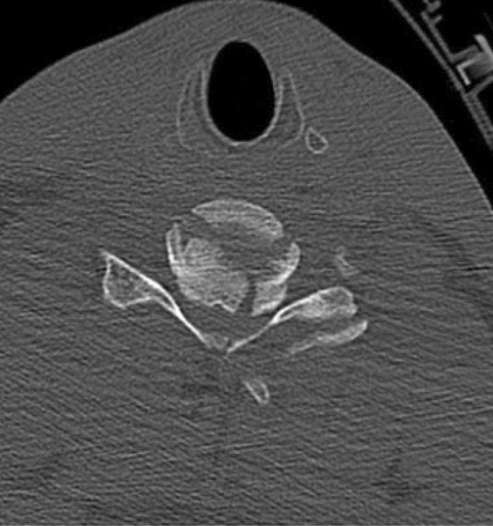
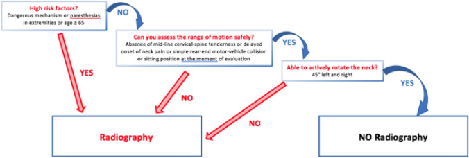
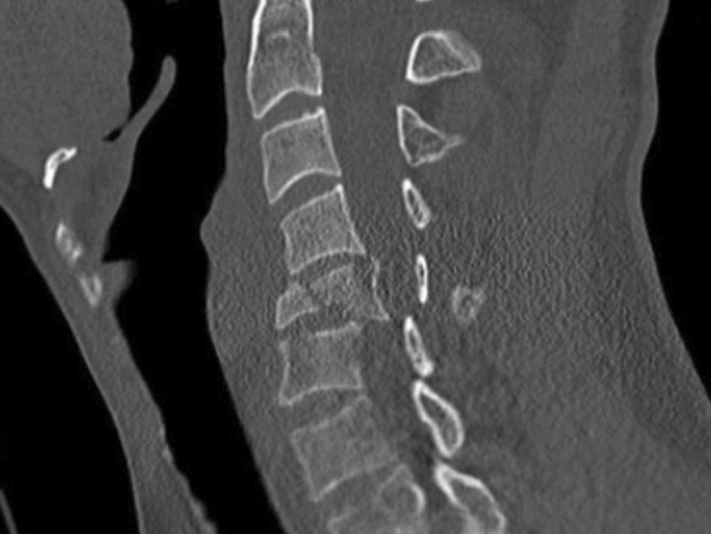
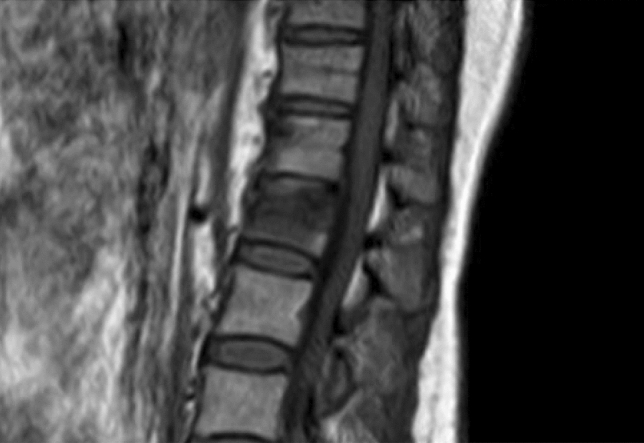
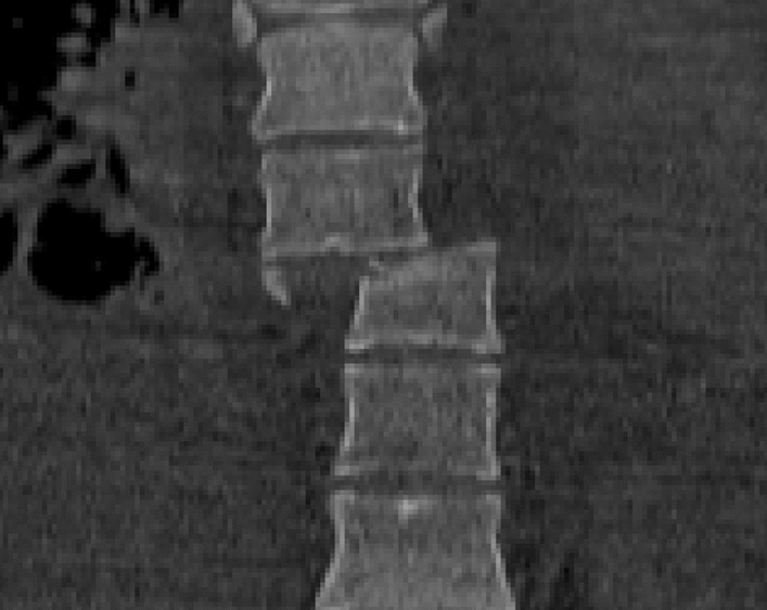
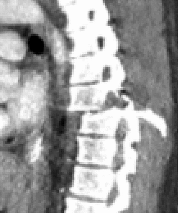
Spinal trauma is an important cause of disability worldwide. Injury to the cervical spine (CS) occurs frequently after major trauma. 5-10% of patients with blunt trauma have a cervical spine injury. The cervical spine accounts for ~ 50% of all spinal injuries. Determination of CS stability is a common challenge in the acute care setting of patients with trauma. Several issues, indeed, are of particular concern: who needs CS imaging; what imaging should be obtained; when should computed tomography (CT), magnetic resonance imaging (MRI), or flexion/extension (F/E) radiographs be obtained; and how is significant ligamentous injury excluded in the comatose patient. CT and MRI both have roles to play. This article aims to present the different imaging to frame techniques to be used with greater precision in the acute event also for the purpose of planning the next therapeutic process. An overview of the applicability of the same methods in forensic pathology is also provided highlighting possible future biomarker to ease in diagnosis of acute TBI.
脊髓创伤是全球致残的一个重要原因。颈椎(CS)损伤在严重创伤后经常发生。5-10%的钝性创伤患者有颈椎损伤。CS 约占所有脊柱损伤的 50%。在创伤患者的急性护理环境中,CS 稳定性的确定是一个常见的挑战。确实有几个问题特别值得关注:谁需要 CS 成像;应该获得哪些影像学检查;何时应进行计算机断层扫描(CT)、磁共振成像(MRI)或屈伸(F/E)射线照相;以及如何在昏迷患者中排除重要的韧带损伤。CT 和 MRI 都有其作用。本文旨在介绍不同的成像方法,以便在急性事件中更精确地使用,也为规划下一个治疗过程。还概述了同一方法在法医病理学中的适用性,突出了可能有助于急性 TBI 诊断的未来生物标志物。