Leiden Institute of Physics, Leiden, The Netherlands.
Biomed Microdevices. 2012 Aug;14(4):641-9. doi: 10.1007/s10544-012-9642-y.
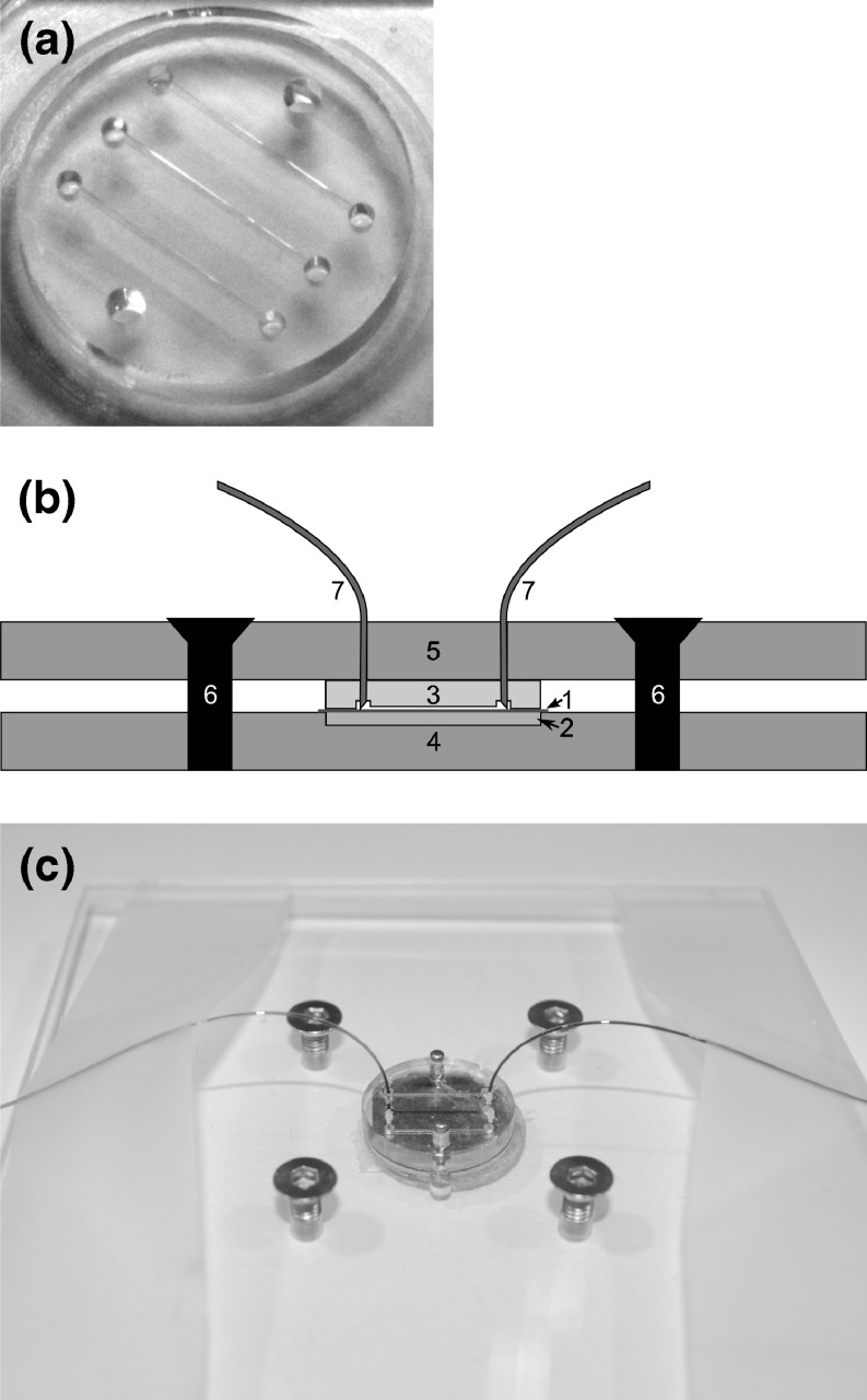
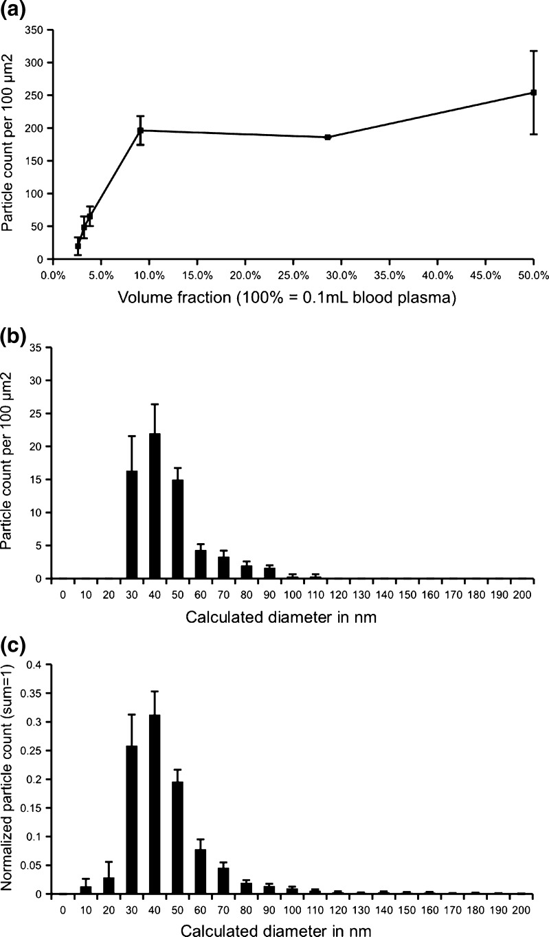
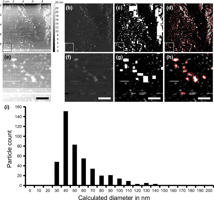
Microparticles, also known as microvesicles, found in blood plasma, urine, and most other body fluids, may serve as valuable biomarkers of diseases such as cardiovascular diseases, systemic inflammatory disease, thrombosis, and cancer. Unfortunately, the detection and quantification of microparticles are hampered by the microscopic size of these particles and their relatively low abundance in blood plasma. The use of a combination of microfluidics and atomic force microscopy to detect microparticles in blood plasma circumvents both problems. In this study, capture of a specific subset of microparticles directly from blood plasma on antibody-coated mica surface is demonstrated. The described method excludes isolation and washing steps to prepare microparticles, improves the detection sensitivity, and yields the size distribution of the captured particles. The majority of the captured particles have a size ranging from 30 to 90 nm, which is in good agreement with prior results obtained with microparticles immediately isolated from fresh plasma. Furthermore, the qualitative shape of the size distribution of microparticles is shown not to be affected by high-speed centrifugation or the use of the microfluidic circuit, demonstrating the relative stable nature of microparticles ex vivo.
微粒,也称为微泡,存在于血浆、尿液和大多数其他体液中,可能是心血管疾病、全身性炎症性疾病、血栓形成和癌症等疾病的有价值的生物标志物。不幸的是,由于这些颗粒的微观尺寸及其在血浆中的相对低丰度,微粒的检测和定量受到阻碍。使用微流控和原子力显微镜的组合来检测血浆中的微粒可以解决这两个问题。在这项研究中,直接从涂有抗体的云母表面从血浆中捕获特定亚群的微粒。所描述的方法排除了用于制备微粒的分离和洗涤步骤,提高了检测灵敏度,并得到了捕获颗粒的尺寸分布。捕获的大多数颗粒的尺寸范围为 30 至 90nm,这与从新鲜血浆中立即分离出的微粒获得的先前结果非常吻合。此外,还表明微粒的尺寸分布的定性形状不受高速离心或微流控电路的影响,证明了微粒在体外的相对稳定性质。