Neurosurgical Department of Affiliated Zhongshan Hospital, Xiamen University, Xiamen, People's Republic of China.
Int J Nanomedicine. 2012;7:1031-41. doi: 10.2147/IJN.S26541. Epub 2012 Feb 23.
Nanobiotechnology can provide more efficient tools for diagnosis, targeted and personalized therapy, and increase the chances of brain tumor treatment being successful. Use of nanoparticles is a promising strategy for overcoming the blood-brain barrier and delivering drugs to the brain. Gelatin-siloxane (GS) nanoparticles modified with Tat peptide can enhance plasmid DNA transfection efficiency compared with a commercial reagent.
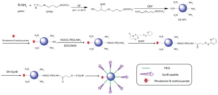
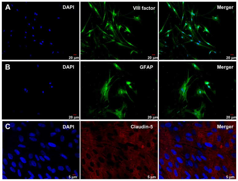
SynB-PEG-GS nanoparticles are membrane-penetrable, and can cross the blood-brain barrier and deliver a drug to its target site in the brain. The efficiency of delivery was investigated in vivo and in vitro using brain capillary endothelial cells, a cocultured blood-brain barrier model, and a normal mouse model.
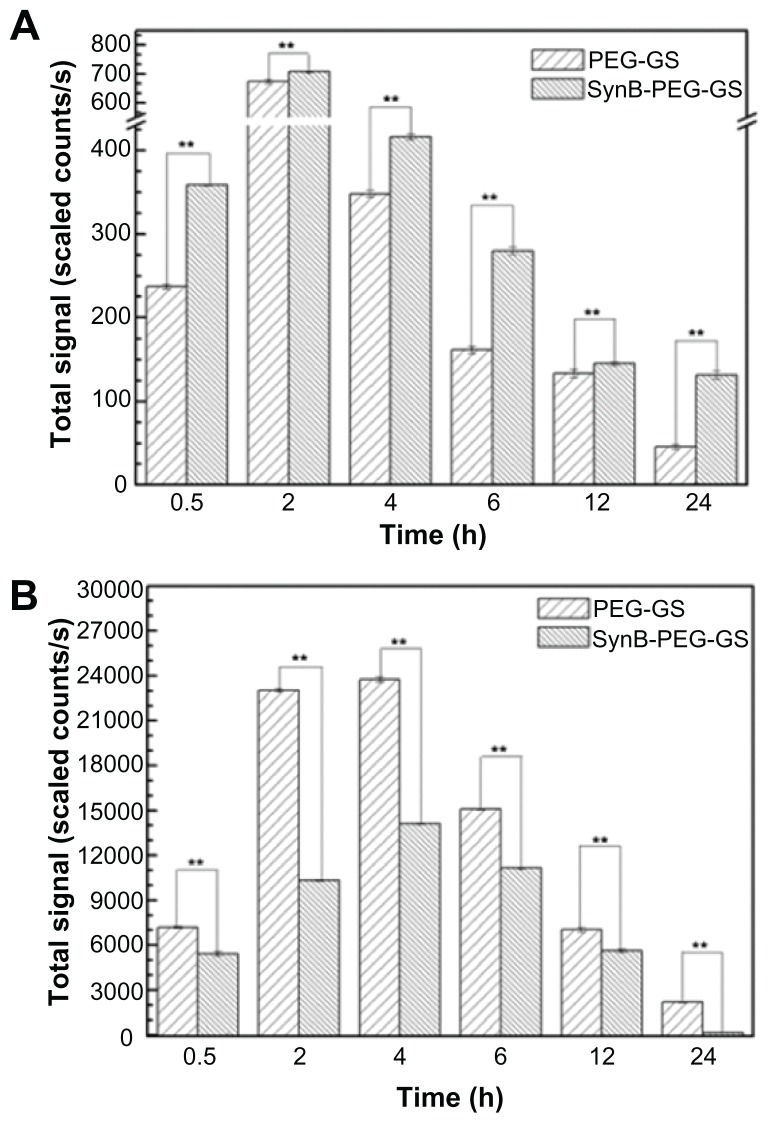
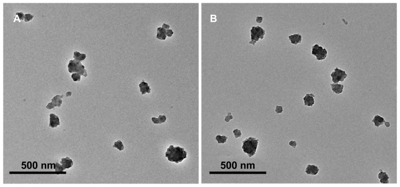
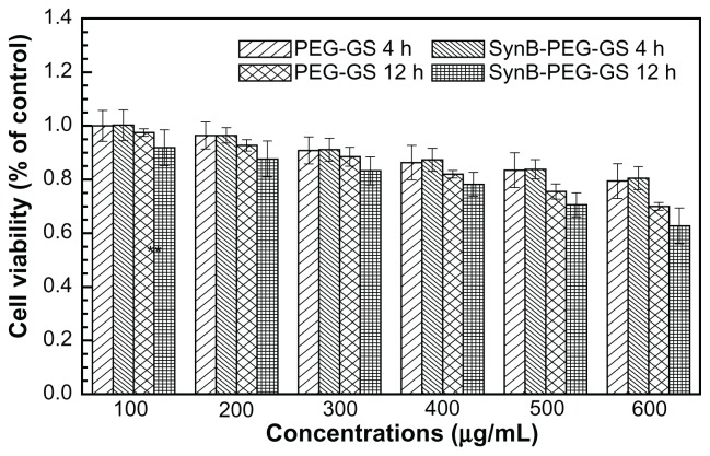
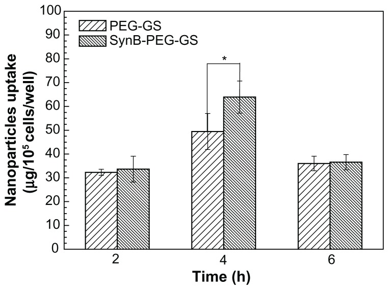
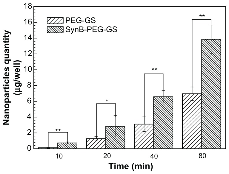
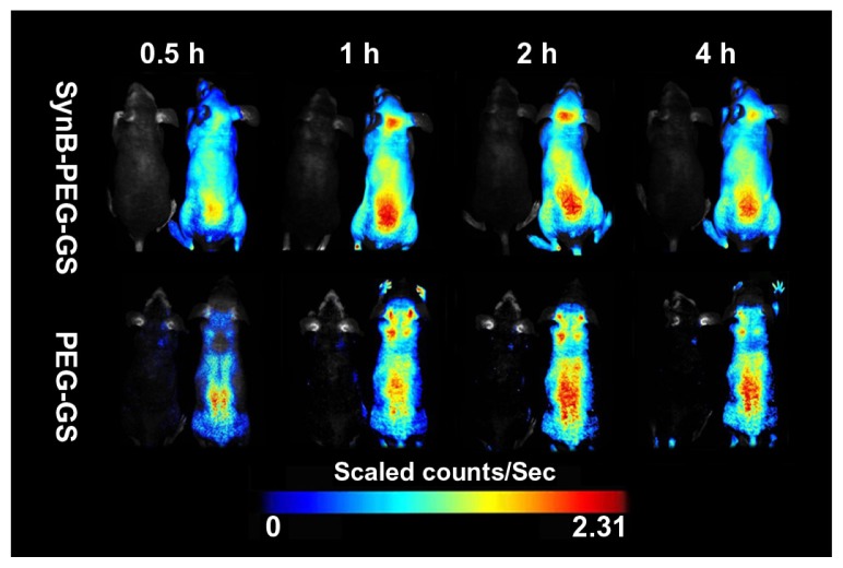
Our study demonstrated that both SynB-PEG-GS and PEG-GS nanoparticles had a spherical shape and an average diameter of 150-200 nm. It was shown by MTT assay that SynB-PEG-GS nanoparticles had good biocompatibility with brain capillary endothelial cells. Cellular uptake by SynB-PEG-GS nanoparticles was higher than that for PEG-GS nanoparticles for all incubation periods. The amount of SynB-PEG-GS nanoparticles crossing the cocultured blood-brain barrier model was significantly higher than that of PEG-GS nanoparticles at all time points measured (P < 0.05). In animal testing, SynB-PEG-GS nanoparticle levels in the brain were significantly higher than those of PEG-GS nanoparticles at all time points measured (P < 0.01). In contrast with localization in the brain, PEG-GS nanoparticle levels were significantly higher than those of SynB-PEG-GS nanoparticles (P < 0.01) in the liver.
This study indicates that SynB-PEG-GS nanoparticles have favorable properties with regard to morphology, size distribution, and toxicity. Moreover, the SynB-PEG-GS nanoparticles exhibited more efficient brain capillary endothelial cell uptake and improved crossing of the blood-brain barrier. Further, biodistribution studies of rhodamine-loaded nanoparticles demonstrated that modification with the SynB peptide could not only improve the ability of PEG-GS nanoparticles to evade capture in the reticuloendothelial system but also enhance their efficiency in crossing the blood-brain barrier.
纳米生物技术可以为诊断、靶向和个性化治疗提供更有效的工具,并增加脑肿瘤治疗成功的机会。使用纳米颗粒是克服血脑屏障并将药物递送到大脑的有前途的策略。用 Tat 肽修饰的明胶-硅氧烷 (GS) 纳米颗粒可以提高质粒 DNA 的转染效率,优于商业试剂。
SynB-PEG-GS 纳米颗粒具有膜穿透性,可以穿过血脑屏障并将药物递送到大脑中的靶部位。在体内和体外使用脑毛细血管内皮细胞、共培养的血脑屏障模型和正常小鼠模型研究了递送效率。
我们的研究表明,SynB-PEG-GS 和 PEG-GS 纳米颗粒均具有球形形状和 150-200nm 的平均直径。MTT 测定表明,SynB-PEG-GS 纳米颗粒与脑毛细血管内皮细胞具有良好的生物相容性。SynB-PEG-GS 纳米颗粒的细胞摄取率在所有孵育期均高于 PEG-GS 纳米颗粒。在所有测量的时间点,穿过共培养的血脑屏障模型的 SynB-PEG-GS 纳米颗粒的量均明显高于 PEG-GS 纳米颗粒(P<0.05)。在动物试验中,在所有测量的时间点,SynB-PEG-GS 纳米颗粒在脑中的水平均明显高于 PEG-GS 纳米颗粒(P<0.01)。与脑内定位相反,PEG-GS 纳米颗粒的水平在肝脏中明显高于 SynB-PEG-GS 纳米颗粒(P<0.01)。
本研究表明,SynB-PEG-GS 纳米颗粒具有形态、大小分布和毒性方面的有利性质。此外,SynB-PEG-GS 纳米颗粒表现出更有效的脑毛细血管内皮细胞摄取和改善血脑屏障通透性。此外,负载罗丹明的纳米颗粒的生物分布研究表明,SynB 肽的修饰不仅可以提高 PEG-GS 纳米颗粒逃避网状内皮系统捕获的能力,还可以提高其穿过血脑屏障的效率。