Bone and Cartilage Research Unit, Institute of pathology, CHU Sart-Tilman, 4000 Liège, Belgium.
Arthritis Res Ther. 2012 Mar 12;14(2):R58. doi: 10.1186/ar3771.
This work aimed at comparing the production of inflammatory and pro- and anti-angiogenic factors by normal/reactive (N/R) or inflammatory (I) areas of the osteoarthritic synovial membrane. The effects of interleukin (IL)-1β and chondroitin sulfate (CS) on the expression of pro- and anti-angiogenic factors by synovial fibroblasts cells (SFC) were also studied.
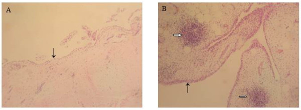
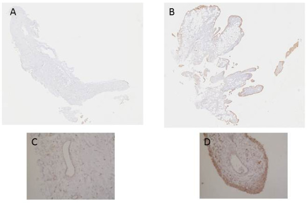
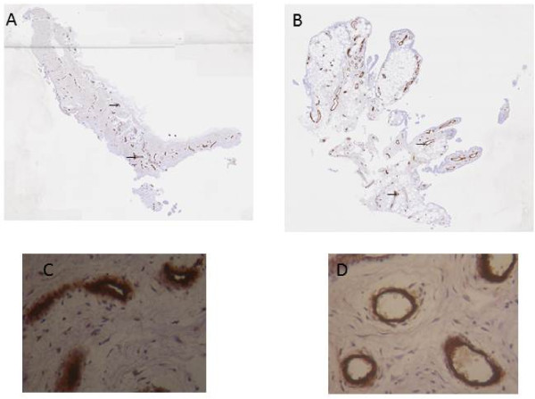
Biopsies from N/R or from I areas of osteoarthritic synovial membrane were collected at the time of surgery. The inflammatory status of the synovial membrane was characterized by the surgeon according to macroscopic criteria, including the synovial vascularization, the villi formation and the hypertrophic aspect of the tissue. We assessed the expression of CD45, von Willebrand factor and vascular endothelial growth factor (VEGF) antigen by immunohistochemistry in both N/R and I biopsies. The production of IL-6, -8, VEGF and thrombospondin (TSP)-1 by N/R or I synovial cells was quantified by ELISA. SFC were cultured in the absence or in the presence of IL-1β (1 ng/ml) and with or without CS (10, 50, 200 μg/ml). Gene expression of pro-angiogenic factors (VEGF, basic fibroblast growth factor (bFGF), nerve growth factor (NGF), matrix metalloproteinase (MMP)-2 and angiopoietin (ang)-1) and anti-angiogenic factors (vascular endothelial growth inhibitor (VEGI), TSP-1 and -2) were determined by real time RT-PCR. Production of VEGI and TSP-1 was also estimated by ELISA.
Immunohistochemistry showed the increase of lymphocyte infiltration, vascular density and VEGF expression in I compared to N/R synovial biopsies. Synovial cells from I areas produced more IL-6, IL-8 and VEGF but less TSP-1 than cells isolated from N/R synovial biopsies. The expression of pro-angiogenic factors by SFC was stimulated by IL-1β. A time dependent regulation of the expression of anti-angiogenic factor genes was observed. IL-1β stimulated the expression of anti-angiogenic factor genes but inhibited it after 24 h. CS reversed the inhibitory effect of IL-1β on anti-angiogenic factors, VEGI and TSP-1.
We demonstrated that synovial biopsies from I areas expressed a pro-angiogenic phenotype. IL-1β induced an imbalance between pro- and anti-angiogenic factors in SFC and CS tended to normalize this IL-1β-induced imbalance, providing a new possible mechanism of action of this drug.
本研究旨在比较骨关节炎滑膜正常/反应性(N/R)或炎症(I)区域产生炎症和促血管生成及抗血管生成因子的情况。还研究了白细胞介素(IL)-1β和硫酸软骨素(CS)对滑膜成纤维细胞(SFC)中促血管生成和抗血管生成因子表达的影响。
在手术时收集 N/R 或骨关节炎滑膜 I 区的活检组织。根据外科医生的宏观标准评估滑膜的炎症状态,包括滑膜血管化、绒毛形成和组织的肥大。我们通过免疫组织化学法评估 N/R 和 I 活检组织中 CD45、血管假性血友病因子和血管内皮生长因子(VEGF)抗原的表达。通过 ELISA 定量检测 N/R 或 I 滑膜细胞产生的 IL-6、-8、VEGF 和血栓调节蛋白(TSP)-1。在无 IL-1β(1ng/ml)或存在 IL-1β(1ng/ml)以及有无 CS(10、50、200μg/ml)的情况下培养 SFC。通过实时 RT-PCR 确定促血管生成因子(VEGF、碱性成纤维细胞生长因子(bFGF)、神经生长因子(NGF)、基质金属蛋白酶(MMP)-2 和血管生成素(ang)-1)和抗血管生成因子(血管内皮生长抑制剂(VEGI)、TSP-1 和 -2)的基因表达。通过 ELISA 还估计了 VEGI 和 TSP-1 的产生。
与 N/R 滑膜活检相比,免疫组织化学显示 I 区淋巴细胞浸润、血管密度和 VEGF 表达增加。I 区滑膜细胞产生的 IL-6、IL-8 和 VEGF 多于 N/R 滑膜细胞,但产生的 TSP-1 较少。SFC 的促血管生成因子表达受 IL-1β刺激。观察到抗血管生成因子基因表达的时间依赖性调节。IL-1β刺激抗血管生成因子基因的表达,但在 24 小时后抑制其表达。CS 逆转了 IL-1β 对抗血管生成因子、VEGI 和 TSP-1 的抑制作用。
我们证明 I 区的滑膜活检表达了促血管生成表型。IL-1β诱导 SFC 中促血管生成和抗血管生成因子之间的失衡,CS 倾向于使这种 IL-1β 诱导的失衡正常化,为这种药物提供了一种新的可能作用机制。