Cellular Neurobiology, Zoological Institute, TU Braunschweig, Braunschweig, Germany.
PLoS One. 2012;7(3):e34167. doi: 10.1371/journal.pone.0034167. Epub 2012 Mar 28.
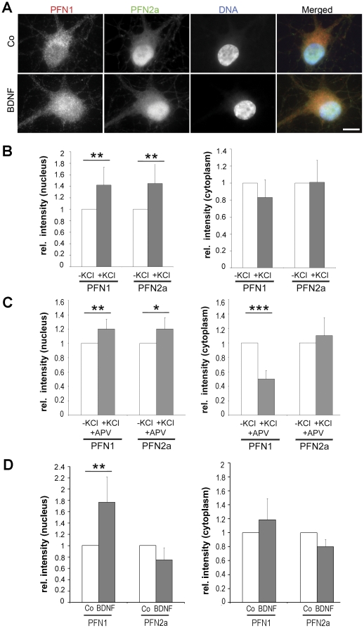
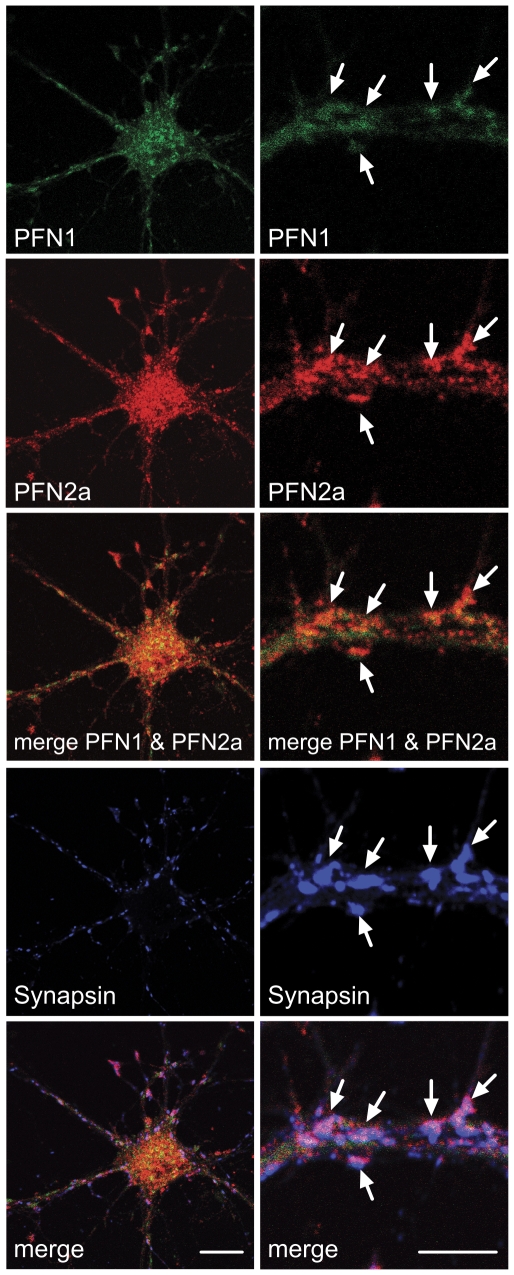
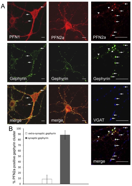
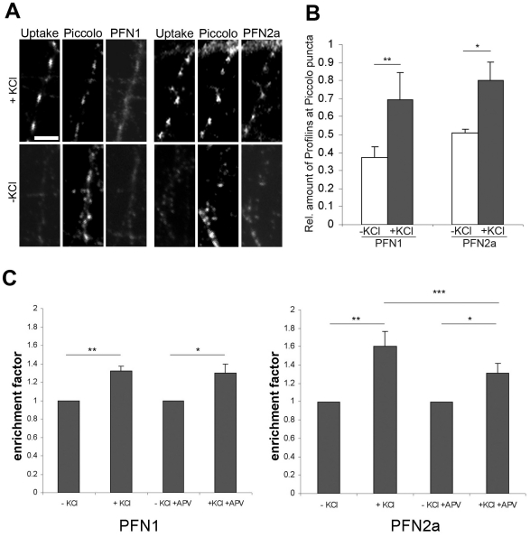
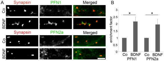
Profilins are prominent regulators of actin dynamics. While most mammalian cells express only one profilin, two isoforms, PFN1 and PFN2a are present in the CNS. To challenge the hypothesis that the expression of two profilin isoforms is linked to the complex shape of neurons and to the activity-dependent structural plasticity, we analysed how PFN1 and PFN2a respond to changes of neuronal activity. Simultaneous labelling of rodent embryonic neurons with isoform-specific monoclonal antibodies revealed both isoforms in the same synapse. Immunoelectron microscopy on brain sections demonstrated both profilins in synapses of the mature rodent cortex, hippocampus and cerebellum. Both isoforms were significantly more abundant in postsynaptic than in presynaptic structures. Immunofluorescence showed PFN2a associated with gephyrin clusters of the postsynaptic active zone in inhibitory synapses of embryonic neurons. When cultures were stimulated in order to change their activity level, active synapses that were identified by the uptake of synaptotagmin antibodies, displayed significantly higher amounts of both isoforms than non-stimulated controls. Specific inhibition of NMDA receptors by the antagonist APV in cultured rat hippocampal neurons resulted in a decrease of PFN2a but left PFN1 unaffected. Stimulation by the brain derived neurotrophic factor (BDNF), on the other hand, led to a significant increase in both synaptic PFN1 and PFN2a. Analogous results were obtained for neuronal nuclei: both isoforms were localized in the same nucleus, and their levels rose significantly in response to KCl stimulation, whereas BDNF caused here a higher increase in PFN1 than in PFN2a. Our results strongly support the notion of an isoform specific role for profilins as regulators of actin dynamics in different signalling pathways, in excitatory as well as in inhibitory synapses. Furthermore, they suggest a functional role for both profilins in neuronal nuclei.
原肌球蛋白是肌动蛋白动态的主要调节因子。虽然大多数哺乳动物细胞只表达一种原肌球蛋白,但在中枢神经系统中存在两种同工型,PFN1 和 PFN2a。为了验证两种原肌球蛋白同工型的表达与神经元的复杂形状和活动依赖性结构可塑性相关的假设,我们分析了 PFN1 和 PFN2a 如何响应神经元活动的变化。用同工型特异性单克隆抗体同时标记啮齿动物胚胎神经元,揭示了两种同工型都存在于同一个突触中。对脑切片进行免疫电子显微镜研究表明,两种原肌球蛋白同工型都存在于成熟啮齿动物皮质、海马体和小脑的突触中。两种同工型在后突触结构中的丰度明显高于前突触结构。免疫荧光显示 PFN2a 与胚胎神经元抑制性突触后活跃区的神经胶质蛋白簇相关。当培养物受到刺激以改变其活动水平时,通过突触结合蛋白抗体摄取来识别的活跃突触显示出两种同工型的含量明显高于未受刺激的对照物。在培养的大鼠海马神经元中,NMDA 受体的拮抗剂 APV 特异性抑制导致 PFN2a 减少,但 PFN1 不受影响。另一方面,脑源性神经营养因子 (BDNF) 的刺激导致突触 PFN1 和 PFN2a 明显增加。对于神经元核也得到了类似的结果:两种同工型都定位于同一个核中,它们的水平在响应 KCl 刺激时显著升高,而 BDNF 在这里导致 PFN1 的增加高于 PFN2a。我们的结果强烈支持原肌球蛋白同工型特异性作为不同信号通路中肌动蛋白动态调节剂的作用的观点,在兴奋性和抑制性突触中都是如此。此外,它们还表明两种原肌球蛋白同工型在神经元核中都具有功能作用。