• 文献检索
  • 文档翻译
  • 深度研究
  • 学术资讯
  • Suppr Zotero 插件Zotero 插件
  • 邀请有礼
  • 套餐&价格
  • 历史记录
应用&插件
Suppr Zotero 插件Zotero 插件浏览器插件Mac 客户端Windows 客户端微信小程序
定价
高级版会员购买积分包购买API积分包
服务
文献检索文档翻译深度研究API 文档MCP 服务
关于我们
关于 Suppr公司介绍联系我们用户协议隐私条款
关注我们

Suppr 超能文献

核心技术专利:CN118964589B侵权必究
粤ICP备2023148730 号-1Suppr @ 2026

文献检索

告别复杂PubMed语法,用中文像聊天一样搜索,搜遍4000万医学文献。AI智能推荐,让科研检索更轻松。

立即免费搜索

文件翻译

保留排版,准确专业,支持PDF/Word/PPT等文件格式,支持 12+语言互译。

免费翻译文档

深度研究

AI帮你快速写综述,25分钟生成高质量综述,智能提取关键信息,辅助科研写作。

立即免费体验

使用CompuCell3D对组织进行多尺度建模。

Multi-scale modeling of tissues using CompuCell3D.

作者信息

Swat Maciej H, Thomas Gilberto L, Belmonte Julio M, Shirinifard Abbas, Hmeljak Dimitrij, Glazier James A

机构信息

Department of Physics, Biocomplexity Institute, Indiana University, Bloomington, Indiana, USA.

出版信息

Methods Cell Biol. 2012;110:325-66. doi: 10.1016/B978-0-12-388403-9.00013-8.

DOI:10.1016/B978-0-12-388403-9.00013-8
PMID:22482955
原文链接:https://pmc.ncbi.nlm.nih.gov/articles/PMC3612985/
Abstract

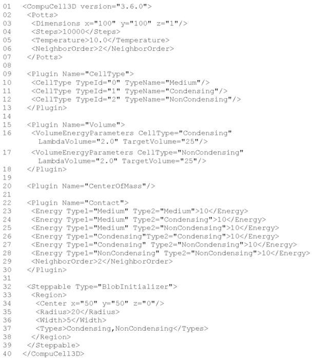
The study of how cells interact to produce tissue development, homeostasis, or diseases was, until recently, almost purely experimental. Now, multi-cell computer simulation methods, ranging from relatively simple cellular automata to complex immersed-boundary and finite-element mechanistic models, allow in silico study of multi-cell phenomena at the tissue scale based on biologically observed cell behaviors and interactions such as movement, adhesion, growth, death, mitosis, secretion of chemicals, chemotaxis, etc. This tutorial introduces the lattice-based Glazier-Graner-Hogeweg (GGH) Monte Carlo multi-cell modeling and the open-source GGH-based CompuCell3D simulation environment that allows rapid and intuitive modeling and simulation of cellular and multi-cellular behaviors in the context of tissue formation and subsequent dynamics. We also present a walkthrough of four biological models and their associated simulations that demonstrate the capabilities of the GGH and CompuCell3D.

摘要

直到最近,关于细胞如何相互作用以产生组织发育、体内平衡或疾病的研究几乎完全是实验性的。现在,从相对简单的细胞自动机到复杂的浸入边界和有限元机制模型等多细胞计算机模拟方法,使得基于生物学观察到的细胞行为和相互作用(如运动、黏附、生长、死亡、有丝分裂、化学物质分泌、趋化性等),在计算机上对组织尺度的多细胞现象进行研究成为可能。本教程介绍基于格点的格拉齐尔 - 格拉纳 - 霍赫韦格(GGH)蒙特卡洛多细胞建模以及基于GGH的开源CompuCell3D模拟环境,该环境允许在组织形成及后续动态变化的背景下,快速直观地对细胞和多细胞行为进行建模与模拟。我们还展示了四个生物学模型及其相关模拟的演练过程,这些演练展示了GGH和CompuCell3D的功能。

https://cdn.ncbi.nlm.nih.gov/pmc/blobs/0961/3612985/635455eb5c7b/nihms438274f34.jpg
https://cdn.ncbi.nlm.nih.gov/pmc/blobs/0961/3612985/8b9e8ca83f71/nihms438274f1.jpg
https://cdn.ncbi.nlm.nih.gov/pmc/blobs/0961/3612985/7804135c5111/nihms438274f2.jpg
https://cdn.ncbi.nlm.nih.gov/pmc/blobs/0961/3612985/faf9691630be/nihms438274f3.jpg
https://cdn.ncbi.nlm.nih.gov/pmc/blobs/0961/3612985/006314772da6/nihms438274f4.jpg
https://cdn.ncbi.nlm.nih.gov/pmc/blobs/0961/3612985/78d82a718f6d/nihms438274f5.jpg
https://cdn.ncbi.nlm.nih.gov/pmc/blobs/0961/3612985/2fef2fa4f59a/nihms438274f6.jpg
https://cdn.ncbi.nlm.nih.gov/pmc/blobs/0961/3612985/010b326586e3/nihms438274f7.jpg
https://cdn.ncbi.nlm.nih.gov/pmc/blobs/0961/3612985/2c6f8274bb2d/nihms438274f8.jpg
https://cdn.ncbi.nlm.nih.gov/pmc/blobs/0961/3612985/5d72c692e1bf/nihms438274f9.jpg
https://cdn.ncbi.nlm.nih.gov/pmc/blobs/0961/3612985/9ee1ba994a50/nihms438274f10.jpg
https://cdn.ncbi.nlm.nih.gov/pmc/blobs/0961/3612985/6205628afce6/nihms438274f11.jpg
https://cdn.ncbi.nlm.nih.gov/pmc/blobs/0961/3612985/7e7b95ac63f2/nihms438274f12.jpg
https://cdn.ncbi.nlm.nih.gov/pmc/blobs/0961/3612985/6c4c2fd74d00/nihms438274f13.jpg
https://cdn.ncbi.nlm.nih.gov/pmc/blobs/0961/3612985/8b36d89ea4ec/nihms438274f14.jpg
https://cdn.ncbi.nlm.nih.gov/pmc/blobs/0961/3612985/f0616596cbb8/nihms438274f15.jpg
https://cdn.ncbi.nlm.nih.gov/pmc/blobs/0961/3612985/8b649aa40b69/nihms438274f16.jpg
https://cdn.ncbi.nlm.nih.gov/pmc/blobs/0961/3612985/0da798eeb1b3/nihms438274f17.jpg
https://cdn.ncbi.nlm.nih.gov/pmc/blobs/0961/3612985/512066900ca5/nihms438274f18.jpg
https://cdn.ncbi.nlm.nih.gov/pmc/blobs/0961/3612985/4144ecc509ac/nihms438274f19.jpg
https://cdn.ncbi.nlm.nih.gov/pmc/blobs/0961/3612985/4d90704da050/nihms438274f20.jpg
https://cdn.ncbi.nlm.nih.gov/pmc/blobs/0961/3612985/bea7a9196068/nihms438274f21.jpg
https://cdn.ncbi.nlm.nih.gov/pmc/blobs/0961/3612985/97f78a5e0423/nihms438274f22a.jpg
https://cdn.ncbi.nlm.nih.gov/pmc/blobs/0961/3612985/5b858feba86a/nihms438274f23.jpg
https://cdn.ncbi.nlm.nih.gov/pmc/blobs/0961/3612985/501e50cae8dc/nihms438274f24.jpg
https://cdn.ncbi.nlm.nih.gov/pmc/blobs/0961/3612985/f60192d05bad/nihms438274f25.jpg
https://cdn.ncbi.nlm.nih.gov/pmc/blobs/0961/3612985/6d5fb9ddf757/nihms438274f26.jpg
https://cdn.ncbi.nlm.nih.gov/pmc/blobs/0961/3612985/ba7485a5db98/nihms438274f27a.jpg
https://cdn.ncbi.nlm.nih.gov/pmc/blobs/0961/3612985/1b1a23881147/nihms438274f28a.jpg
https://cdn.ncbi.nlm.nih.gov/pmc/blobs/0961/3612985/da710d2c1934/nihms438274f29.jpg
https://cdn.ncbi.nlm.nih.gov/pmc/blobs/0961/3612985/3f93181b5c8c/nihms438274f30.jpg
https://cdn.ncbi.nlm.nih.gov/pmc/blobs/0961/3612985/69ec1be664d0/nihms438274f31.jpg
https://cdn.ncbi.nlm.nih.gov/pmc/blobs/0961/3612985/e5be18625428/nihms438274f32.jpg
https://cdn.ncbi.nlm.nih.gov/pmc/blobs/0961/3612985/494ce2e25a42/nihms438274f33.jpg
https://cdn.ncbi.nlm.nih.gov/pmc/blobs/0961/3612985/635455eb5c7b/nihms438274f34.jpg

相似文献

1
Multi-scale modeling of tissues using CompuCell3D.使用CompuCell3D对组织进行多尺度建模。
Methods Cell Biol. 2012;110:325-66. doi: 10.1016/B978-0-12-388403-9.00013-8.
2
Simulation of single-species bacterial-biofilm growth using the Glazier-Graner-Hogeweg model and the CompuCell3D modeling environment.使用Glazier-Graner-Hogeweg模型和CompuCell3D建模环境对单物种细菌生物膜生长进行模拟。
Math Biosci Eng. 2008 Apr;5(2):355-88. doi: 10.3934/mbe.2008.5.355.
3
Multicell simulations of development and disease using the CompuCell3D simulation environment.使用CompuCell3D模拟环境对发育和疾病进行多细胞模拟。
Methods Mol Biol. 2009;500:361-428. doi: 10.1007/978-1-59745-525-1_13.
4
Integrating intracellular dynamics using CompuCell3D and Bionetsolver: applications to multiscale modelling of cancer cell growth and invasion.使用 CompuCell3D 和 Bionetsolver 整合细胞内动力学:在癌症细胞生长和入侵的多尺度建模中的应用。
PLoS One. 2012;7(3):e33726. doi: 10.1371/journal.pone.0033726. Epub 2012 Mar 26.
5
Space-Limited Mitosis in the Glazier-Graner-Hogeweg Model.格拉齐尔-格拉纳-霍赫韦格模型中的空间受限有丝分裂
Bull Math Biol. 2017 Jan;79(1):1-20. doi: 10.1007/s11538-016-0204-y. Epub 2016 Nov 1.
6
Development of a coupled simulation toolkit for computational radiation biology based on Geant4 and CompuCell3D.基于 Geant4 和 CompuCell3D 的计算放射生物学耦合模拟工具包的开发。
Phys Med Biol. 2021 Feb 11;66(4):045026. doi: 10.1088/1361-6560/abd4f9.
7
Cell-based multi-parametric model of cleft progression during submandibular salivary gland branching morphogenesis.基于细胞的下颌下腺分支形态发生过程中裂隙进展的多参数模型。
PLoS Comput Biol. 2013;9(11):e1003319. doi: 10.1371/journal.pcbi.1003319. Epub 2013 Nov 21.
8
A computational model predicting disruption of blood vessel development.一种预测血管发育中断的计算模型。
PLoS Comput Biol. 2013 Apr;9(4):e1002996. doi: 10.1371/journal.pcbi.1002996. Epub 2013 Apr 4.
9
Computer simulations of cell sorting due to differential adhesion.基于细胞黏附差异的细胞分选计算机模拟
PLoS One. 2011;6(10):e24999. doi: 10.1371/journal.pone.0024999. Epub 2011 Oct 18.
10
Computer simulation of cellular patterning within the Drosophila pupal eye.果蝇蛹眼内细胞模式形成的计算机模拟。
PLoS Comput Biol. 2010 Jul 1;6(7):e1000841. doi: 10.1371/journal.pcbi.1000841.

引用本文的文献

1
Computational modeling and simulation in oncology.肿瘤学中的计算建模与模拟
Clin Transl Med. 2025 Sep;15(9):e70456. doi: 10.1002/ctm2.70456.
2
Fluid-derived lattices for unbiased modeling of bacterial colony growth.用于细菌菌落生长无偏建模的流体衍生晶格
PLoS One. 2025 Aug 28;20(8):e0330491. doi: 10.1371/journal.pone.0330491. eCollection 2025.
3
V-Cornea: A computational model of corneal epithelium homeostasis, injury, and recovery.V-角膜:角膜上皮稳态、损伤与恢复的计算模型。

本文引用的文献

1
Modelling Morphogenesis: From Single Cells to Crawling Slugs.形态发生建模:从单细胞到爬行蛞蝓
J Theor Biol. 1997 Feb 7;184(3):229-235. doi: 10.1006/jtbi.1996.0237.
2
Computer simulations of cell sorting due to differential adhesion.基于细胞黏附差异的细胞分选计算机模拟
PLoS One. 2011;6(10):e24999. doi: 10.1371/journal.pone.0024999. Epub 2011 Oct 18.
3
A multi-cell, multi-scale model of vertebrate segmentation and somite formation.脊椎动物分节和体节形成的多细胞多尺度模型。
bioRxiv. 2025 Aug 14:2025.08.11.669602. doi: 10.1101/2025.08.11.669602.
4
Temporal dynamics of angiogenesis: the emerging role of mechanoregulated pathways.血管生成的时间动态变化:机械调节通路的新作用
Biochem Soc Trans. 2025 Aug 29;53(4):909-923. doi: 10.1042/BST20253048.
5
Radiobiological Modeling with Monte Carlo Tools - Simulating Cellular Responses to Ionizing Radiation.使用蒙特卡洛工具进行放射生物学建模——模拟细胞对电离辐射的反应。
Technol Cancer Res Treat. 2025 Jan-Dec;24:15330338251350909. doi: 10.1177/15330338251350909. Epub 2025 Jul 17.
6
Shape matters: inferring the motility of confluent cells from static images.形状很重要:从静态图像推断汇合细胞的运动性。
Soft Matter. 2025 Jul 7. doi: 10.1039/d5sm00222b.
7
Personalizing computational models to construct medical digital twins.个性化计算模型以构建医学数字孪生体。
J R Soc Interface. 2025 Jul;22(228):20250055. doi: 10.1098/rsif.2025.0055. Epub 2025 Jul 2.
8
Proliferation and regeneration of the healthy human urothelium: A multi-scale simulation approach with 16 hypotheses of cell differentiation.健康人膀胱尿路上皮的增殖与再生:一种具有16种细胞分化假设的多尺度模拟方法。
PLoS One. 2025 Jun 20;20(6):e0325132. doi: 10.1371/journal.pone.0325132. eCollection 2025.
9
Review of the geometrical developments in GEANT4-DNA: From a biological perspective.从生物学角度对GEANT4-DNA中几何发展的综述。
Rev Phys. 2025 Dec;13. doi: 10.1016/j.revip.2025.100110. Epub 2025 Feb 11.
10
Galvanotactic directionality of cell groups depends on group size.细胞群的电趋性方向性取决于群体大小。
Proc Natl Acad Sci U S A. 2025 May 27;122(21):e2416440122. doi: 10.1073/pnas.2416440122. Epub 2025 May 20.
PLoS Comput Biol. 2011 Oct;7(10):e1002155. doi: 10.1371/journal.pcbi.1002155. Epub 2011 Oct 6.
4
3D multi-cell simulation of tumor growth and angiogenesis.三维多细胞肿瘤生长和血管生成模拟。
PLoS One. 2009 Oct 16;4(10):e7190. doi: 10.1371/journal.pone.0007190.
5
Dynamic mechanisms of blood vessel growth.血管生长的动态机制。
Nonlinearity. 2006;19(1):C1-C10. doi: 10.1088/0951-7715/19/1/000.
6
From Genes to Organisms Via the Cell A Problem-Solving Environment for Multicellular Development.从基因到经由细胞的生物体 一个用于多细胞发育的问题解决环境
Comput Sci Eng. 2007;9(4):50-60. doi: 10.1109/MCSE.2007.74.
7
Contact-inhibited chemotaxis in de novo and sprouting blood-vessel growth.在新生血管生成和血管芽生过程中的接触抑制趋化作用。
PLoS Comput Biol. 2008 Sep 19;4(9):e1000163. doi: 10.1371/journal.pcbi.1000163.
8
Simulation of single-species bacterial-biofilm growth using the Glazier-Graner-Hogeweg model and the CompuCell3D modeling environment.使用Glazier-Graner-Hogeweg模型和CompuCell3D建模环境对单物种细菌生物膜生长进行模拟。
Math Biosci Eng. 2008 Apr;5(2):355-88. doi: 10.3934/mbe.2008.5.355.
9
Adhesion between cells, diffusion of growth factors, and elasticity of the AER produce the paddle shape of the chick limb.细胞间的黏附、生长因子的扩散以及顶外胚层嵴(AER)的弹性造就了鸡胚肢体的桨状形态。
Physica A. 2007 Jan 1;373:521-532. doi: 10.1016/j.physa.2006.05.028.
10
Coordinated action of N-CAM, N-cadherin, EphA4, and ephrinB2 translates genetic prepatterns into structure during somitogenesis in chick.在鸡胚体节形成过程中,N-CAM、N-钙黏蛋白、EphA4和ephrinB2的协同作用将遗传预模式转化为结构。
Curr Top Dev Biol. 2008;81:205-47. doi: 10.1016/S0070-2153(07)81007-6.