• 文献检索
  • 文档翻译
  • 深度研究
  • 学术资讯
  • Suppr Zotero 插件Zotero 插件
  • 邀请有礼
  • 套餐&价格
  • 历史记录
应用&插件
Suppr Zotero 插件Zotero 插件浏览器插件Mac 客户端Windows 客户端微信小程序
定价
高级版会员购买积分包购买API积分包
服务
文献检索文档翻译深度研究API 文档MCP 服务
关于我们
关于 Suppr公司介绍联系我们用户协议隐私条款
关注我们

Suppr 超能文献

核心技术专利:CN118964589B侵权必究
粤ICP备2023148730 号-1Suppr @ 2026

文献检索

告别复杂PubMed语法,用中文像聊天一样搜索,搜遍4000万医学文献。AI智能推荐,让科研检索更轻松。

立即免费搜索

文件翻译

保留排版,准确专业,支持PDF/Word/PPT等文件格式,支持 12+语言互译。

免费翻译文档

深度研究

AI帮你快速写综述,25分钟生成高质量综述,智能提取关键信息,辅助科研写作。

立即免费体验

活体追踪生物发光鼠疫耶尔森菌对腺鼠疫动态的成像。

Imaging of bubonic plague dynamics by in vivo tracking of bioluminescent Yersinia pestis.

机构信息

Yersinia Research Unit, Institut Pasteur, Paris, France.

出版信息

PLoS One. 2012;7(4):e34714. doi: 10.1371/journal.pone.0034714. Epub 2012 Apr 5.

DOI:10.1371/journal.pone.0034714
PMID:22496846
原文链接:https://pmc.ncbi.nlm.nih.gov/articles/PMC3320629/
Abstract

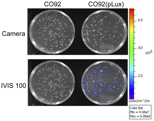
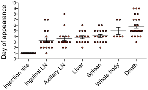
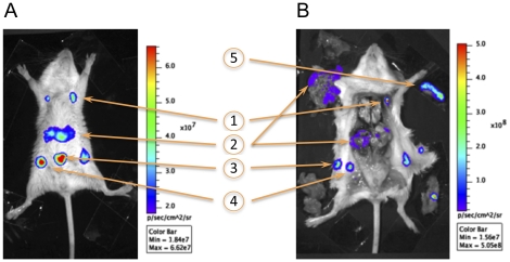
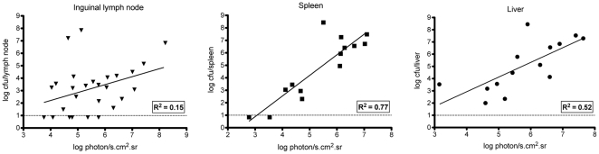
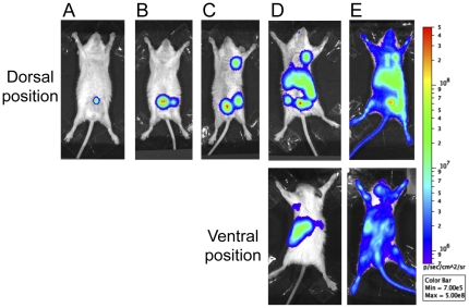
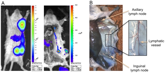
Yersinia pestis dissemination in a host is usually studied by enumerating bacteria in the tissues of animals sacrificed at different times. This laborious methodology gives only snapshots of the infection, as the infectious process is not synchronized. In this work we used in vivo bioluminescence imaging (BLI) to follow Y. pestis dissemination during bubonic plague. We first demonstrated that Y. pestis CO92 transformed with pGEN-luxCDABE stably emitted bioluminescence in vitro and in vivo, while retaining full virulence. The light produced from live animals allowed to delineate the infected organs and correlated with bacterial loads, thus validating the BLI tool. We then showed that the first step of the infectious process is a bacterial multiplication at the injection site (linea alba), followed by a colonization of the draining inguinal lymph node(s), and subsequently of the ipsilateral axillary lymph node through a direct connection between the two nodes. A mild bacteremia and an effective filtering of the blood stream by the liver and spleen probably accounted for the early bacterial blood clearance and the simultaneous development of bacterial foci within these organs. The saturation of the filtering capacity of the spleen and liver subsequently led to terminal septicemia. Our results also indicate that secondary lymphoid tissues are the main targets of Y. pestis multiplication and that colonization of other organs occurs essentially at the terminal phase of the disease. Finally, our analysis reveals that the high variability in the kinetics of infection is attributable to the time the bacteria remain confined at the injection site. However, once Y. pestis has reached the draining lymph nodes, the disease progresses extremely rapidly, leading to the invasion of the entire body within two days and to death of the animals. This highlights the extraordinary capacity of Y. pestis to annihilate the host innate immune response.

摘要

鼠疫耶尔森氏菌在宿主中的传播通常通过对不同时间处死的动物组织中的细菌进行计数来研究。这种费力的方法只能给出感染的快照,因为感染过程不同步。在这项工作中,我们使用体内生物发光成像(BLI)来跟踪鼠疫耶尔森氏菌在腺鼠疫中的传播。我们首先证明,用 pGEN-luxCDABE 转化的鼠疫耶尔森氏菌 CO92 在体外和体内稳定地发出生物发光,同时保持完全的毒力。来自活体动物的光允许描绘感染的器官,并与细菌负荷相关联,从而验证了 BLI 工具。然后我们表明,感染过程的第一步是在注射部位(白线)细菌繁殖,随后是引流腹股沟淋巴结的定植,然后是同侧腋窝淋巴结的定植,通过两个淋巴结之间的直接连接。轻度菌血症和肝脏和脾脏对血流的有效过滤可能导致早期细菌清除和这些器官内细菌灶的同时发展。脾脏和肝脏过滤能力的饱和随后导致终末期败血症。我们的结果还表明,次级淋巴组织是鼠疫耶尔森氏菌繁殖的主要靶标,而其他器官的定植主要发生在疾病的终末期。最后,我们的分析表明,感染动力学的高度可变性归因于细菌在注射部位被限制的时间。然而,一旦鼠疫耶尔森氏菌到达引流淋巴结,疾病就会迅速进展,导致两天内全身感染并导致动物死亡。这突出了鼠疫耶尔森氏菌消灭宿主先天免疫反应的非凡能力。

https://cdn.ncbi.nlm.nih.gov/pmc/blobs/112a/3320629/e5001433c7c5/pone.0034714.g006.jpg
https://cdn.ncbi.nlm.nih.gov/pmc/blobs/112a/3320629/36b6ca13d47d/pone.0034714.g001.jpg
https://cdn.ncbi.nlm.nih.gov/pmc/blobs/112a/3320629/4649ef989360/pone.0034714.g002.jpg
https://cdn.ncbi.nlm.nih.gov/pmc/blobs/112a/3320629/04687e2e3ea5/pone.0034714.g003.jpg
https://cdn.ncbi.nlm.nih.gov/pmc/blobs/112a/3320629/cd891f298773/pone.0034714.g004.jpg
https://cdn.ncbi.nlm.nih.gov/pmc/blobs/112a/3320629/753317b505a2/pone.0034714.g005.jpg
https://cdn.ncbi.nlm.nih.gov/pmc/blobs/112a/3320629/e5001433c7c5/pone.0034714.g006.jpg

相似文献

1
Imaging of bubonic plague dynamics by in vivo tracking of bioluminescent Yersinia pestis.活体追踪生物发光鼠疫耶尔森菌对腺鼠疫动态的成像。
PLoS One. 2012;7(4):e34714. doi: 10.1371/journal.pone.0034714. Epub 2012 Apr 5.
2
Bioluminescent tracing of a Yersinia pestis pCD1-mutant and Yersinia pseudotuberculosis in subcutaneously infected mice.生物发光示踪鼠疫耶尔森氏菌 pCD1 突变株和假结核耶尔森氏菌在皮下感染小鼠中的作用。
Microbes Infect. 2018 Mar;20(3):166-175. doi: 10.1016/j.micinf.2017.11.005. Epub 2017 Nov 26.
3
Bioluminescent tracking of colonization and clearance dynamics of plasmid-deficient Yersinia pestis strains in a mouse model of septicemic plague.生物发光示踪法研究质粒缺陷型鼠疫耶尔森菌在败血症型鼠疫小鼠模型中的定殖和清除动力学。
Microbes Infect. 2014 Mar;16(3):214-24. doi: 10.1016/j.micinf.2013.11.013. Epub 2013 Dec 11.
4
Development of bioluminescent bioreporters for in vitro and in vivo tracking of Yersinia pestis.用于鼠疫耶尔森氏菌体外和体内追踪的生物发光生物报告器的开发。
PLoS One. 2012;7(10):e47123. doi: 10.1371/journal.pone.0047123. Epub 2012 Oct 11.
5
Bioluminescence imaging to track bacterial dissemination of Yersinia pestis using different routes of infection in mice.生物发光成像技术用于追踪鼠疫耶尔森氏菌在小鼠体内不同感染途径的传播。
BMC Microbiol. 2012 Jul 24;12:147. doi: 10.1186/1471-2180-12-147.
6
Yersinia pestis subverts the dermal neutrophil response in a mouse model of bubonic plague.鼠疫耶尔森菌在小鼠腺鼠疫模型中颠覆了皮肤中性粒细胞的反应。
mBio. 2013 Aug 27;4(5):e00170-13. doi: 10.1128/mBio.00170-13.
7
Yersinia pestis YopJ suppresses tumor necrosis factor alpha induction and contributes to apoptosis of immune cells in the lymph node but is not required for virulence in a rat model of bubonic plague.鼠疫耶尔森菌的YopJ蛋白可抑制肿瘤坏死因子α的诱导产生,并促使淋巴结中的免疫细胞发生凋亡,但在腺鼠疫大鼠模型中,它并非毒力所必需。
Infect Immun. 2006 Sep;74(9):5126-31. doi: 10.1128/IAI.00219-06.
8
Dissociation of Tissue Destruction and Bacterial Expansion during Bubonic Plague.腺鼠疫期间组织破坏与细菌扩散的分离
PLoS Pathog. 2015 Oct 20;11(10):e1005222. doi: 10.1371/journal.ppat.1005222. eCollection 2015 Oct.
9
A non-invasive in vivo imaging system to study dissemination of bioluminescent Yersinia pestis CO92 in a mouse model of pneumonic plague.一种用于研究生物发光鼠疫耶尔森菌 CO92 在肺炎型鼠疫小鼠模型中传播的非侵入性体内成像系统。
Microb Pathog. 2013 Feb;55:39-50. doi: 10.1016/j.micpath.2012.09.011. Epub 2012 Oct 9.
10
Transcriptomic and innate immune responses to Yersinia pestis in the lymph node during bubonic plague.鼠疫耶尔森菌在腹股沟淋巴结中引起的转录组和固有免疫反应。
Infect Immun. 2010 Dec;78(12):5086-98. doi: 10.1128/IAI.00256-10. Epub 2010 Sep 27.

引用本文的文献

1
Plague history, Mongol history, and the processes of focalisation leading up to the Black Death: a response to Brack .鼠疫史、蒙古史以及导致黑死病的聚焦过程:对布拉克的回应
Med Hist. 2025 Feb 17;68(4):1-25. doi: 10.1017/mdh.2024.29.
2
The physiological landscape and specificity of antibody repertoires are consolidated by multiple immunizations.抗体库的生理格局和特异性通过多次免疫得以巩固。
Elife. 2024 Dec 18;13:e92718. doi: 10.7554/eLife.92718.
3
JCM1395 Achieved Intratumoral Growth with Minimal Inflammation: Evidence for Live Bacterial Therapeutic Potential by an Optimized Sample Preparation and Colony PCR Method.

本文引用的文献

1
Early systemic bacterial dissemination and a rapid innate immune response characterize genetic resistance to plague of SEG mice.早期系统性细菌播散和快速先天免疫反应是 SEG 鼠对鼠疫遗传抗性的特征。
J Infect Dis. 2012 Jan 1;205(1):134-43. doi: 10.1093/infdis/jir696. Epub 2011 Nov 16.
2
Investigation of a researcher's death due to septicemic plague.一名研究人员因败血性鼠疫死亡的调查。
N Engl J Med. 2011 Jun 30;364(26):2563-4. doi: 10.1056/NEJMc1010939.
3
Monitoring of gene expression in bacteria during infections using an adaptable set of bioluminescent, fluorescent and colorigenic fusion vectors.
JCM1395实现瘤内生长且炎症轻微:通过优化样本制备和菌落PCR方法证明活细菌的治疗潜力
Pharmaceutics. 2023 Mar 19;15(3):989. doi: 10.3390/pharmaceutics15030989.
4
The invasive pathogen Yersinia pestis disrupts host blood vasculature to spread and provoke hemorrhages.侵袭性病原体鼠疫耶尔森菌破坏宿主血管系统以传播并引发出血。
PLoS Negl Trop Dis. 2021 Oct 5;15(10):e0009832. doi: 10.1371/journal.pntd.0009832. eCollection 2021 Oct.
5
Comparison of the transmission efficiency and plague progression dynamics associated with two mechanisms by which fleas transmit Yersinia pestis.比较两种跳蚤传播鼠疫耶尔森菌的机制的传播效率和鼠疫进展动力学。
PLoS Pathog. 2020 Dec 7;16(12):e1009092. doi: 10.1371/journal.ppat.1009092. eCollection 2020 Dec.
6
The Genome of the Great Gerbil Reveals Species-Specific Duplication of an MHCII Gene.大沙鼠基因组揭示 MHCII 基因的种特异性重复。
Genome Biol Evol. 2020 Feb 1;12(2):3832-3849. doi: 10.1093/gbe/evaa008.
7
Longitudinal bioluminescent imaging of HIV-1 infection during antiretroviral therapy and treatment interruption in humanized mice.抗逆转录病毒治疗和治疗中断期间人源化小鼠 HIV-1 感染的纵向生物发光成像。
PLoS Pathog. 2019 Dec 5;15(12):e1008161. doi: 10.1371/journal.ppat.1008161. eCollection 2019 Dec.
8
Metabolomic analyses reveal lipid abnormalities and hepatic dysfunction in non-human primate model for Yersinia pestis.代谢组学分析揭示了鼠疫耶尔森菌非人灵长类动物模型中的脂质异常和肝功能障碍。
Metabolomics. 2018 Dec 29;15(1):2. doi: 10.1007/s11306-018-1457-2.
9
Enterobacteria and host resistance to infection.肠杆菌与宿主抗感染抵抗力
Mamm Genome. 2018 Aug;29(7-8):558-576. doi: 10.1007/s00335-018-9749-4. Epub 2018 May 21.
10
A Bioluminescent Francisella tularensis SCHU S4 Strain Enables Noninvasive Tracking of Bacterial Dissemination and the Evaluation of Antibiotics in an Inhalational Mouse Model of Tularemia.一种生物发光的土拉弗朗西斯菌SCHU S4菌株能够在土拉菌病吸入性小鼠模型中对细菌传播进行非侵入性追踪并评估抗生素。
Antimicrob Agents Chemother. 2016 Nov 21;60(12):7206-7215. doi: 10.1128/AAC.01586-16. Print 2016 Dec.
利用一组适应性强的生物发光、荧光和显色融合载体监测感染过程中细菌的基因表达。
PLoS One. 2011;6(6):e20425. doi: 10.1371/journal.pone.0020425. Epub 2011 Jun 3.
4
Novel genetic tools for diaminopimelic acid selection in virulence studies of Yersinia pestis.新型遗传工具用于鼠疫耶尔森氏菌毒力研究中的二氨基庚二酸选择。
PLoS One. 2011 Mar 2;6(3):e17352. doi: 10.1371/journal.pone.0017352.
5
Emerging infectious disease: what are the relative roles of ecology and evolution?新发传染病:生态与进化的相对作用是什么?
Trends Ecol Evol. 1995 Aug;10(8):319-24. doi: 10.1016/s0169-5347(00)89118-1.
6
In vivo analysis of Yersinia enterocolitica infection using luxCDABE.利用 luxCDABE 进行肠侵袭性大肠杆菌感染的活体分析。
FEMS Microbiol Lett. 2010 Jun;307(2):201-6. doi: 10.1111/j.1574-6968.2010.01983.x.
7
Construction of a bioluminescence reporter plasmid for Francisella tularensis.构建一株弗氏志贺菌生物发光报告质粒。
Plasmid. 2010 Nov;64(3):156-61. doi: 10.1016/j.plasmid.2010.07.001. Epub 2010 Jul 8.
8
Evaluation of biophotonic imaging to estimate bacterial burden in mice infected with highly virulent compared to less virulent Streptococcus pneumoniae serotypes.评价生物光子学成像技术在估计高毒力与低毒力肺炎链球菌血清型感染小鼠细菌负荷中的应用。
Antimicrob Agents Chemother. 2010 Aug;54(8):3155-60. doi: 10.1128/AAC.00310-10. Epub 2010 Jun 7.
9
Optimisation of bioluminescent reporters for use with mycobacteria.用于分枝杆菌的生物发光报告基因的优化。
PLoS One. 2010 May 24;5(5):e10777. doi: 10.1371/journal.pone.0010777.
10
The search for early markers of plague: evidence for accumulation of soluble Yersinia pestis LcrV in bubonic and pneumonic mouse models of disease.鼠疫早期标志物的探寻:在腺鼠疫和肺鼠疫小鼠疾病模型中可溶性鼠疫耶尔森菌LcrV积累的证据
FEMS Immunol Med Microbiol. 2010 Jul 1;59(2):197-206. doi: 10.1111/j.1574-695X.2010.00687.x. Epub 2010 Apr 19.