• 文献检索
  • 文档翻译
  • 深度研究
  • 学术资讯
  • Suppr Zotero 插件Zotero 插件
  • 邀请有礼
  • 套餐&价格
  • 历史记录
应用&插件
Suppr Zotero 插件Zotero 插件浏览器插件Mac 客户端Windows 客户端微信小程序
定价
高级版会员购买积分包购买API积分包
服务
文献检索文档翻译深度研究API 文档MCP 服务
关于我们
关于 Suppr公司介绍联系我们用户协议隐私条款
关注我们

Suppr 超能文献

核心技术专利:CN118964589B侵权必究
粤ICP备2023148730 号-1Suppr @ 2026

文献检索

告别复杂PubMed语法,用中文像聊天一样搜索,搜遍4000万医学文献。AI智能推荐,让科研检索更轻松。

立即免费搜索

文件翻译

保留排版,准确专业,支持PDF/Word/PPT等文件格式,支持 12+语言互译。

免费翻译文档

深度研究

AI帮你快速写综述,25分钟生成高质量综述,智能提取关键信息,辅助科研写作。

立即免费体验

一种使用新型三模态 MRI-光声-Raman 纳米粒子的脑肿瘤分子成像策略。

A brain tumor molecular imaging strategy using a new triple-modality MRI-photoacoustic-Raman nanoparticle.

机构信息

Molecular Imaging Program at Stanford, Department of Radiology, Stanford University, California, USA.

出版信息

Nat Med. 2012 Apr 15;18(5):829-34. doi: 10.1038/nm.2721.

DOI:10.1038/nm.2721
PMID:22504484
原文链接:https://pmc.ncbi.nlm.nih.gov/articles/PMC3422133/
Abstract

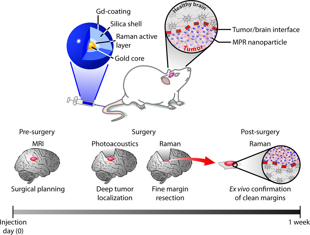
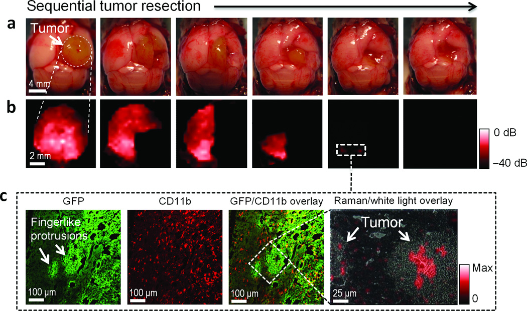
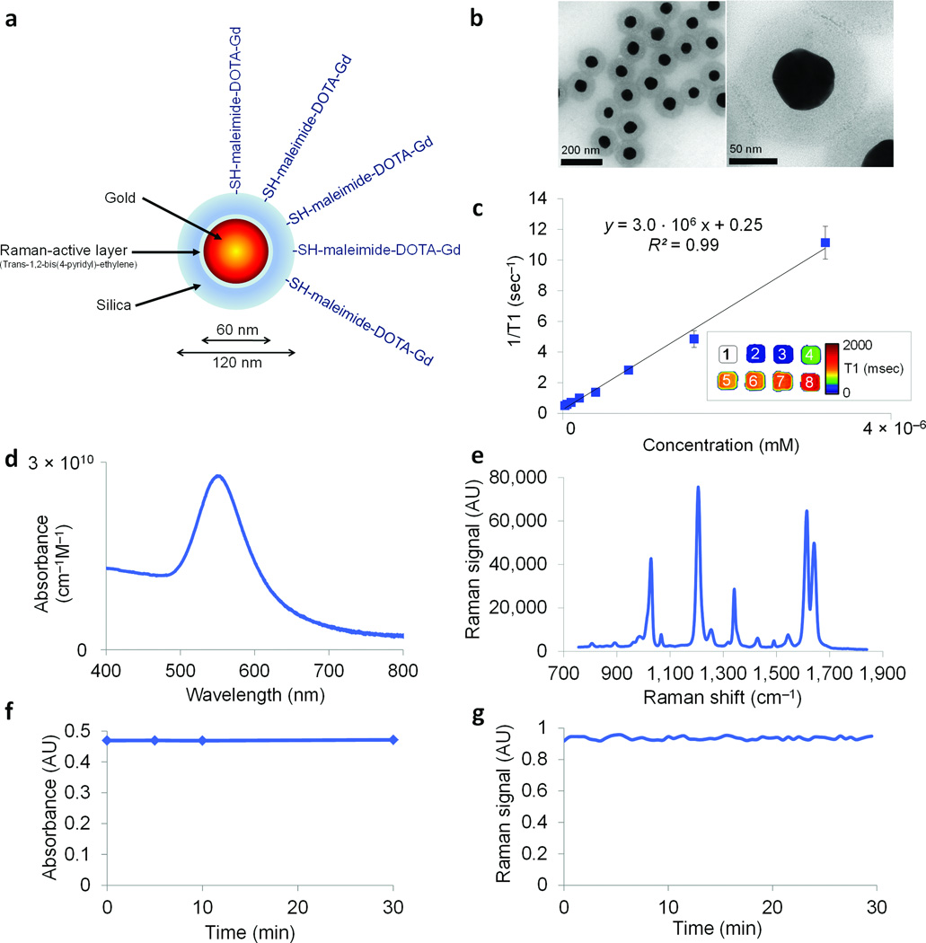
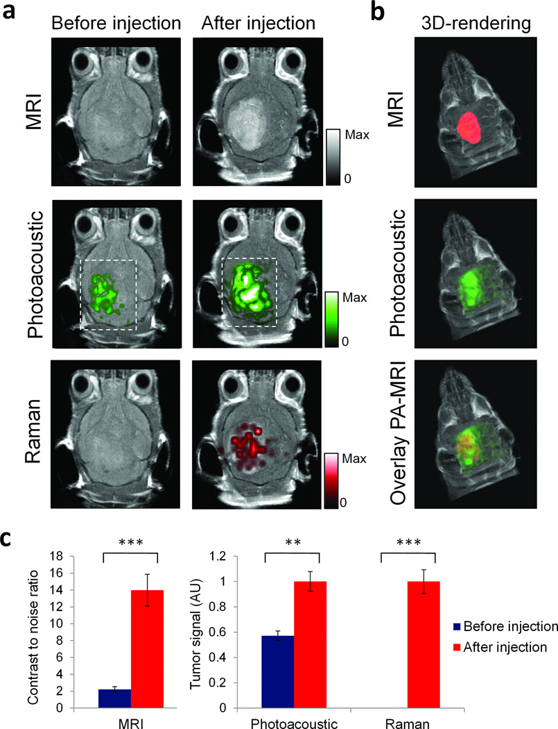
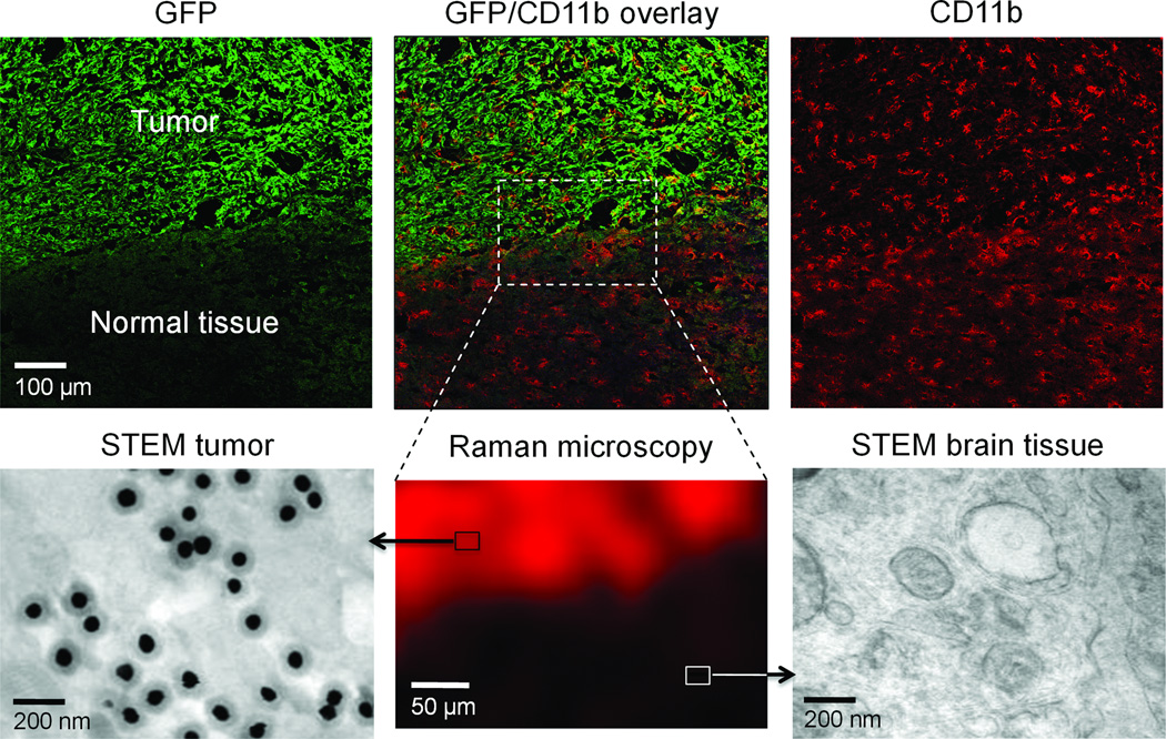
The difficulty in delineating brain tumor margins is a major obstacle in the path toward better outcomes for patients with brain tumors. Current imaging methods are often limited by inadequate sensitivity, specificity and spatial resolution. Here we show that a unique triple-modality magnetic resonance imaging-photoacoustic imaging-Raman imaging nanoparticle (termed here MPR nanoparticle) can accurately help delineate the margins of brain tumors in living mice both preoperatively and intraoperatively. The MPRs were detected by all three modalities with at least a picomolar sensitivity both in vitro and in living mice. Intravenous injection of MPRs into glioblastoma-bearing mice led to MPR accumulation and retention by the tumors, with no MPR accumulation in the surrounding healthy tissue, allowing for a noninvasive tumor delineation using all three modalities through the intact skull. Raman imaging allowed for guidance of intraoperative tumor resection, and a histological correlation validated that Raman imaging was accurately delineating the brain tumor margins. This new triple-modality-nanoparticle approach has promise for enabling more accurate brain tumor imaging and resection.

摘要

脑肿瘤边界的界定困难是提高脑肿瘤患者治疗效果的主要障碍。目前的成像方法通常受到灵敏度、特异性和空间分辨率不足的限制。在这里,我们展示了一种独特的三模态磁共振成像-光声成像-拉曼成像纳米粒子(称为 MPR 纳米粒子),它可以在活体小鼠中准确地帮助术前和术中界定脑肿瘤的边界。在体外和活体小鼠中,MPR 都可以通过所有三种模态进行检测,其灵敏度至少达到皮摩尔级。将 MPR 注入胶质母细胞瘤荷瘤小鼠体内后,MPR 会在肿瘤中积累和保留,而周围健康组织中没有 MPR 积累,从而可以通过完整的颅骨使用所有三种模态进行无创肿瘤界定。拉曼成像可以指导术中肿瘤切除,组织学相关性验证了拉曼成像可以准确地界定脑肿瘤边界。这种新的三模态纳米粒子方法有望实现更精确的脑肿瘤成像和切除。

https://cdn.ncbi.nlm.nih.gov/pmc/blobs/6d9f/3422133/ac6f20f72cc4/nihms331893f5.jpg
https://cdn.ncbi.nlm.nih.gov/pmc/blobs/6d9f/3422133/39bd2894515b/nihms331893f1.jpg
https://cdn.ncbi.nlm.nih.gov/pmc/blobs/6d9f/3422133/cf6e1be81518/nihms331893f2.jpg
https://cdn.ncbi.nlm.nih.gov/pmc/blobs/6d9f/3422133/d5d1b7e30119/nihms331893f3.jpg
https://cdn.ncbi.nlm.nih.gov/pmc/blobs/6d9f/3422133/f50a1b5164f8/nihms331893f4.jpg
https://cdn.ncbi.nlm.nih.gov/pmc/blobs/6d9f/3422133/ac6f20f72cc4/nihms331893f5.jpg

相似文献

1
A brain tumor molecular imaging strategy using a new triple-modality MRI-photoacoustic-Raman nanoparticle.一种使用新型三模态 MRI-光声-Raman 纳米粒子的脑肿瘤分子成像策略。
Nat Med. 2012 Apr 15;18(5):829-34. doi: 10.1038/nm.2721.
2
Dual-Modality Surface-Enhanced Resonance Raman Scattering and Multispectral Optoacoustic Tomography Nanoparticle Approach for Brain Tumor Delineation.双模态表面增强共振拉曼散射和多光谱光声断层成像纳米粒子方法用于脑肿瘤描绘。
Small. 2018 Jun;14(23):e1800740. doi: 10.1002/smll.201800740. Epub 2018 May 4.
3
High Precision Imaging of Microscopic Spread of Glioblastoma with a Targeted Ultrasensitive SERRS Molecular Imaging Probe.使用靶向超灵敏表面增强拉曼散射分子成像探针实现胶质母细胞瘤微观扩散的高精度成像。
Theranostics. 2016 May 7;6(8):1075-84. doi: 10.7150/thno.13842. eCollection 2016.
4
Precise visual distinction of brain glioma from normal tissues via targeted photoacoustic and fluorescence navigation.通过靶向光声和荧光导航,精确区分脑胶质瘤与正常组织。
Nanomedicine. 2020 Jul;27:102204. doi: 10.1016/j.nano.2020.102204. Epub 2020 Apr 12.
5
Surface-Enhanced Resonance Raman Scattering-Guided Brain Tumor Surgery Showing Prognostic Benefit in Rat Models.基于表面增强共振拉曼散射的脑肿瘤手术在大鼠模型中显示出预后获益。
ACS Appl Mater Interfaces. 2019 May 1;11(17):15241-15250. doi: 10.1021/acsami.9b00227. Epub 2019 Apr 17.
6
Gold nanorods for ovarian cancer detection with photoacoustic imaging and resection guidance via Raman imaging in living mice.金纳米棒用于卵巢癌检测,通过活体小鼠的光声成像和拉曼成像进行切除指导。
ACS Nano. 2012 Nov 27;6(11):10366-77. doi: 10.1021/nn304347g. Epub 2012 Nov 2.
7
Non-invasive Imaging of Cancer Using Surface-Enhanced Spatially Offset Raman Spectroscopy (SESORS).使用表面增强空间偏移拉曼光谱学(SESORS)进行癌症的无创成像。
Theranostics. 2019 Aug 13;9(20):5899-5913. doi: 10.7150/thno.36321. eCollection 2019.
8
Photoacoustic signal amplification through plasmonic nanoparticle aggregation.通过等离子体纳米粒子聚集实现光声信号放大。
J Biomed Opt. 2013 Jan;18(1):16001. doi: 10.1117/1.JBO.18.1.016001.
9
Application of multi-wavelength technique for photoacoustic imaging to delineate tumor margins during maximum-safe resection of glioma: A preliminary simulation study.多波长技术在最大安全切除脑胶质瘤过程中的光声成像中的应用:初步模拟研究。
J Clin Neurosci. 2019 Dec;70:242-246. doi: 10.1016/j.jocn.2019.08.040. Epub 2019 Aug 30.
10
Magnetic resonance and photoacoustic imaging of brain tumor mediated by mesenchymal stem cell labeled with multifunctional nanoparticle introduced via carotid artery injection.经颈动脉注射导入多功能纳米颗粒标记的间充质干细胞介导的脑肿瘤磁共振和光声成像。
Nanotechnology. 2018 Apr 20;29(16):165101. doi: 10.1088/1361-6528/aaaf16.

引用本文的文献

1
Oncolytic Therapies for Glioblastoma: Advances, Challenges, and Future Perspectives.胶质母细胞瘤的溶瘤疗法:进展、挑战与未来展望
Cancers (Basel). 2025 Aug 1;17(15):2550. doi: 10.3390/cancers17152550.
2
Applications of magnetic nanoparticles for boundarics in biomedicine.磁性纳米颗粒在生物医学边界中的应用。
Fundam Res. 2025 Jan 2;5(4):1401-1422. doi: 10.1016/j.fmre.2024.12.017. eCollection 2025 Jul.
3
Surface-Enhanced Raman Spectroscopy (SERS) Based Biological and Environmental 2D and 3D Imaging.基于表面增强拉曼光谱(SERS)的生物与环境二维及三维成像

本文引用的文献

1
Advanced contrast nanoagents for photoacoustic molecular imaging, cytometry, blood test and photothermal theranostics.用于光声分子成像、细胞计数、血液检测和光热治疗的先进对比纳米试剂。
Contrast Media Mol Imaging. 2011 Sep-Oct;6(5):346-69. doi: 10.1002/cmmi.455.
2
Noninvasive cell-tracking methods.非侵入式细胞示踪方法。
Nat Rev Clin Oncol. 2011 Sep 27;8(11):677-88. doi: 10.1038/nrclinonc.2011.141.
3
Raman's "effect" on molecular imaging.拉曼对分子成像的影响。
ACS Environ Au. 2025 May 30;5(4):342-362. doi: 10.1021/acsenvironau.4c00149. eCollection 2025 Jul 16.
4
A Permeabilization Workflow To Enable Specific Multiplexed Profiling Using SERS Nanoparticles.一种使用表面增强拉曼散射纳米颗粒实现特定多重分析的透化工作流程。
ACS Appl Mater Interfaces. 2025 Jul 2;17(26):37747-37762. doi: 10.1021/acsami.5c08079. Epub 2025 Jun 17.
5
Artificial intelligence-based radiogenomics reveals the potential immunoregulatory role of COL22A1 in glioma and its induced autoimmune encephalitis.基于人工智能的放射基因组学揭示了COL22A1在胶质瘤及其诱发的自身免疫性脑炎中的潜在免疫调节作用。
Front Immunol. 2025 Mar 6;16:1562070. doi: 10.3389/fimmu.2025.1562070. eCollection 2025.
6
Selective Photothermal Eradication of Glioblastoma Cells Coexisting with Astrocytes by Anti-EGFR-Coated Raman Tags.通过抗表皮生长因子受体(EGFR)包被的拉曼标签选择性光热消除与星形胶质细胞共存的胶质母细胞瘤细胞
ACS Appl Bio Mater. 2025 Apr 21;8(4):3119-3126. doi: 10.1021/acsabm.4c01986. Epub 2025 Mar 18.
7
Nanophotonic-enhanced photoacoustic imaging for brain tumor detection.用于脑肿瘤检测的纳米光子增强光声成像
J Nanobiotechnology. 2025 Mar 5;23(1):170. doi: 10.1186/s12951-025-03204-5.
8
In vivo surface-enhanced Raman scattering techniques: nanoprobes, instrumentation, and applications.体内表面增强拉曼散射技术:纳米探针、仪器设备及应用
Light Sci Appl. 2025 Feb 11;14(1):79. doi: 10.1038/s41377-024-01718-5.
9
"Beyond the Knife"-Applying Theranostic Technologies to Enhance Outcomes in Neurosurgical Oncology.《超越手术刀——应用诊疗技术改善神经外科肿瘤治疗效果》
Brain Sci. 2024 Dec 13;14(12):1253. doi: 10.3390/brainsci14121253.
10
High Spatiotemporal Near-Infrared II Fluorescence Lifetime Imaging for Quantitative Detection of Clinical Tumor Margins.用于临床肿瘤边缘定量检测的高时空近红外II荧光寿命成像
Adv Sci (Weinh). 2025 Feb;12(5):e2411272. doi: 10.1002/advs.202411272. Epub 2024 Dec 9.
J Nucl Med. 2011 Dec;52(12):1839-44. doi: 10.2967/jnumed.111.087775. Epub 2011 Aug 25.
4
Preclinical evaluation of Raman nanoparticle biodistribution for their potential use in clinical endoscopy imaging.用于临床内镜成像的拉曼纳米粒子生物分布的临床前评估。
Small. 2011 Aug 8;7(15):2232-40. doi: 10.1002/smll.201002317. Epub 2011 May 24.
5
The fate and toxicity of Raman-active silica-gold nanoparticles in mice.拉曼活性硅-金纳米粒子在小鼠体内的命运和毒性。
Sci Transl Med. 2011 Apr 20;3(79):79ra33. doi: 10.1126/scitranslmed.3001963.
6
Oxidative stress mediates the effects of Raman-active gold nanoparticles in human cells.氧化应激介导了具有拉曼活性的金纳米粒子在人细胞中的作用。
Small. 2011 Jan 3;7(1):126-36. doi: 10.1002/smll.201001466.
7
Weight of evidence approach for the relative hazard ranking of nanomaterials.基于证据权重的纳米材料相对危害分级方法。
Nanotoxicology. 2011 Sep;5(3):445-58. doi: 10.3109/17435390.2010.512986. Epub 2010 Sep 23.
8
Ex vivo and in vivo diagnosis of C6 glioblastoma development by Raman spectroscopy coupled to a microprobe.通过与微探针耦合的拉曼光谱术对 C6 神经胶质瘤的体外表征和体内诊断。
Anal Bioanal Chem. 2010 Sep;398(1):477-87. doi: 10.1007/s00216-010-3910-6. Epub 2010 Jun 26.
9
Ultrahigh sensitivity carbon nanotube agents for photoacoustic molecular imaging in living mice.用于活体小鼠光声分子成像的超高灵敏度碳纳米管试剂。
Nano Lett. 2010 Jun 9;10(6):2168-72. doi: 10.1021/nl100890d.
10
The phosphatase and tensin homolog regulates epidermal growth factor receptor (EGFR) inhibitor response by targeting EGFR for degradation.磷酸酶和张力蛋白同源物通过靶向 EGFR 进行降解来调节表皮生长因子受体 (EGFR) 抑制剂的反应。
Proc Natl Acad Sci U S A. 2010 Apr 6;107(14):6459-64. doi: 10.1073/pnas.0911188107. Epub 2010 Mar 22.