National ESCA and Surface Analysis Center for Biomedical Problems, University of Washington, Seattle, Washington 98195-1750, United States.
Anal Chem. 2012 Jun 5;84(11):4880-5. doi: 10.1021/ac300480g. Epub 2012 May 11.
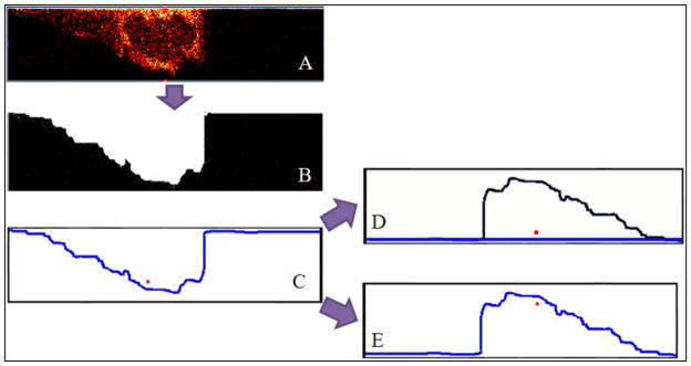
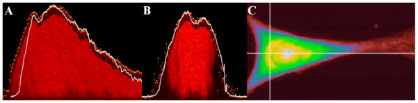
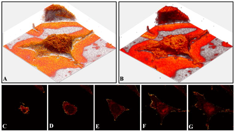
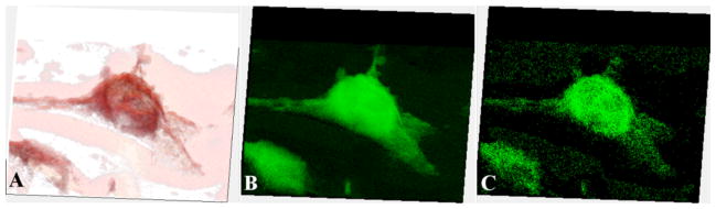
Proper display of three-dimensional time-of-flight secondary ion mass spectrometry (ToF-SIMS) imaging data of complex, nonflat samples requires a correction of the data in the z-direction. Inaccuracies in displaying three-dimensional ToF-SIMS data arise from projecting data from a nonflat surface onto a 2D image plane, as well as possible variations in the sputter rate of the sample being probed. The current study builds on previous studies by creating software written in Matlab, the ZCorrectorGUI (available at http://mvsa.nb.uw.edu/), to apply the z-correction to entire 3D data sets. Three-dimensional image data sets were acquired from NIH/3T3 fibroblasts by collecting ToF-SIMS images, using a dual beam approach (25 keV Bi(3)(+) for analysis cycles and 20 keV C(60)(2+) for sputter cycles). The entire data cube was then corrected by using the new ZCorrectorGUI software, producing accurate chemical information from single cells in 3D. For the first time, a three-dimensional corrected view of a lipid-rich subcellular region, possibly the nuclear membrane, is presented. Additionally, the key assumption of a constant sputter rate throughout the data acquisition was tested by using ToF-SIMS and atomic force microscopy (AFM) analysis of the same cells. For the dried NIH/3T3 fibroblasts examined in this study, the sputter rate was found to not change appreciably in x, y, or z, and the cellular material was sputtered at a rate of approximately 10 nm per 1.25 × 10(13) ions C(60)(2+)/cm(2).
正确显示复杂非平整样品的三维飞行时间二次离子质谱(ToF-SIMS)成像数据需要对 z 方向的数据进行校正。在显示三维 ToF-SIMS 数据时,由于从非平整表面投影数据到 2D 图像平面,以及被探测样品的溅射率可能存在变化,因此会产生不准确。本研究在前人的研究基础上,使用 Matlab 编写软件,即 ZCorrectorGUI(可在 http://mvsa.nb.uw.edu/ 获得),将 z 校正应用于整个 3D 数据集,从而进一步扩展了研究。通过使用双束方法(25keV Bi(3)(+) 进行分析循环和 20keV C(60)(2+) 进行溅射循环),从 NIH/3T3 成纤维细胞采集 ToF-SIMS 图像,获取三维图像数据集。然后,使用新的 ZCorrectorGUI 软件对整个数据立方体进行校正,从 3D 中的单个细胞中获得准确的化学信息。首次呈现了富含脂质的亚细胞区域(可能是核膜)的三维校正视图。此外,还通过对相同细胞进行 ToF-SIMS 和原子力显微镜(AFM)分析,测试了整个数据采集过程中溅射率保持不变的关键假设。对于本研究中检查的干燥 NIH/3T3 成纤维细胞,发现 x、y 和 z 方向的溅射率没有明显变化,细胞材料的溅射速率约为每 1.25×10(13)个 C(60)(2+)离子/cm(2)溅射 10nm。