• 文献检索
  • 文档翻译
  • 深度研究
  • 学术资讯
  • Suppr Zotero 插件Zotero 插件
  • 邀请有礼
  • 套餐&价格
  • 历史记录
应用&插件
Suppr Zotero 插件Zotero 插件浏览器插件Mac 客户端Windows 客户端微信小程序
定价
高级版会员购买积分包购买API积分包
服务
文献检索文档翻译深度研究API 文档MCP 服务
关于我们
关于 Suppr公司介绍联系我们用户协议隐私条款
关注我们

Suppr 超能文献

核心技术专利:CN118964589B侵权必究
粤ICP备2023148730 号-1Suppr @ 2026

文献检索

告别复杂PubMed语法,用中文像聊天一样搜索,搜遍4000万医学文献。AI智能推荐,让科研检索更轻松。

立即免费搜索

文件翻译

保留排版,准确专业,支持PDF/Word/PPT等文件格式,支持 12+语言互译。

免费翻译文档

深度研究

AI帮你快速写综述,25分钟生成高质量综述,智能提取关键信息,辅助科研写作。

立即免费体验

使用光谱域光学相干断层扫描对人体口腔组织进行无创体内结构和血管成像。

Noninvasive in vivo structural and vascular imaging of human oral tissues with spectral domain optical coherence tomography.

作者信息

Davoudi Bahar, Lindenmaier Andras, Standish Beau A, Allo Ghassan, Bizheva Kostadinka, Vitkin Alex

出版信息

Biomed Opt Express. 2012 May 1;3(5):826-39. doi: 10.1364/BOE.3.000826. Epub 2012 Apr 2.

DOI:10.1364/BOE.3.000826
PMID:22567578
原文链接:https://pmc.ncbi.nlm.nih.gov/articles/PMC3342190/
Abstract

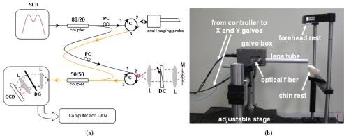
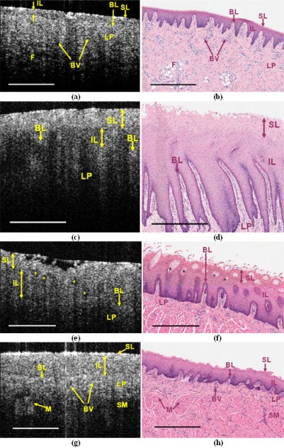
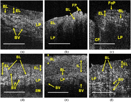
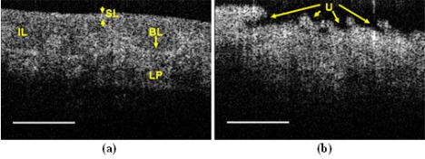
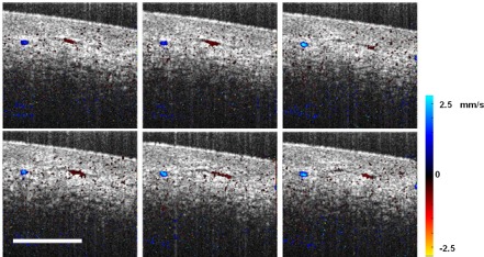
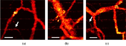
A spectral domain optical coherence tomography (SD-OCT) system and an oral imaging probe have been developed to visualize the microstructural morphology and microvasculature in the human oral cavity. Structural OCT images of ex vivo pig oral tissues with the histology of the same sites were acquired and compared for correlations. Structural in vivo OCT images of healthy human tissue as well as a pathologic site (ulcer) were obtained and analyzed based on the results of the ex vivo pig study, drawing on the similarity between human and swine oral tissues. In vivo Doppler and speckle variance OCT images of the oral cavity in human volunteers were also acquired, to demonstrate the feasibility of microvascular imaging of healthy and pathologic (scar) oral tissue.

摘要

已开发出一种光谱域光学相干断层扫描(SD-OCT)系统和一种口腔成像探头,用于可视化人体口腔中的微观结构形态和微血管系统。采集了离体猪口腔组织相同部位的组织学结构OCT图像,并进行比较以寻找相关性。基于离体猪研究的结果,利用人类和猪口腔组织之间的相似性,获取并分析了健康人体组织以及病理部位(溃疡)的活体结构OCT图像。还采集了人类志愿者口腔的活体多普勒和散斑方差OCT图像,以证明对健康和病理(瘢痕)口腔组织进行微血管成像的可行性。

https://cdn.ncbi.nlm.nih.gov/pmc/blobs/d320/3342190/5919d57016c1/boe-3-5-826-g008.jpg
https://cdn.ncbi.nlm.nih.gov/pmc/blobs/d320/3342190/828a145d2564/boe-3-5-826-g001.jpg
https://cdn.ncbi.nlm.nih.gov/pmc/blobs/d320/3342190/7708966676d6/boe-3-5-826-g002.jpg
https://cdn.ncbi.nlm.nih.gov/pmc/blobs/d320/3342190/38eac5b77580/boe-3-5-826-g003.jpg
https://cdn.ncbi.nlm.nih.gov/pmc/blobs/d320/3342190/b83d09757168/boe-3-5-826-g004.jpg
https://cdn.ncbi.nlm.nih.gov/pmc/blobs/d320/3342190/7f3525feaace/boe-3-5-826-g005.jpg
https://cdn.ncbi.nlm.nih.gov/pmc/blobs/d320/3342190/1ab99cb30e26/boe-3-5-826-g006.jpg
https://cdn.ncbi.nlm.nih.gov/pmc/blobs/d320/3342190/e51db14db482/boe-3-5-826-g007.jpg
https://cdn.ncbi.nlm.nih.gov/pmc/blobs/d320/3342190/5919d57016c1/boe-3-5-826-g008.jpg

相似文献

1
Noninvasive in vivo structural and vascular imaging of human oral tissues with spectral domain optical coherence tomography.使用光谱域光学相干断层扫描对人体口腔组织进行无创体内结构和血管成像。
Biomed Opt Express. 2012 May 1;3(5):826-39. doi: 10.1364/BOE.3.000826. Epub 2012 Apr 2.
2
In vivo imaging of functional microvasculature within tissue beds of oral and nasal cavities by swept-source optical coherence tomography with a forward/side-viewing probe.使用前视/侧视探头的扫频光学相干断层扫描对口腔和鼻腔组织床内的功能性微血管进行体内成像。
Biomed Opt Express. 2014 Jul 15;5(8):2620-34. doi: 10.1364/BOE.5.002620. eCollection 2014 Aug 1.
3
A novel integration of spectral-domain optical-coherence-tomography and laser-ablation system for precision treatment.一种新型的光谱域光学相干层析成像和激光烧蚀系统的集成,用于精确治疗。
Int J Comput Assist Radiol Surg. 2018 Mar;13(3):411-423. doi: 10.1007/s11548-017-1664-8. Epub 2017 Sep 9.
4
Dose-dependent retinal changes following sodium iodate administration: application of spectral-domain optical coherence tomography for monitoring of retinal injury and endogenous regeneration.碘酸钠给药后视网膜的剂量依赖性变化:光谱域光学相干断层扫描在监测视网膜损伤和内源性再生中的应用
Curr Eye Res. 2014 Oct;39(10):1033-41. doi: 10.3109/02713683.2014.892996. Epub 2014 Mar 24.
5
In vivo imaging in the oral cavity by endoscopic optical coherence tomography.内窥式光学相干断层扫描在口腔内的活体成像。
J Biomed Opt. 2018 Mar;23(7):1-13. doi: 10.1117/1.JBO.23.7.071207.
6
Noninvasive structural and microvascular anatomy of oral mucosae using handheld optical coherence tomography.使用手持式光学相干断层扫描技术对口腔黏膜进行无创性结构和微血管解剖
Biomed Opt Express. 2017 Oct 13;8(11):5001-5012. doi: 10.1364/BOE.8.005001. eCollection 2017 Nov 1.
7
Improved speckle contrast optical coherence tomography angiography.改进的散斑对比度光学相干断层扫描血管造影术。
Am J Transl Res. 2018 Oct 15;10(10):3025-3035. eCollection 2018.
8
Microvascular imaging and monitoring of human oral cavity lesions in vivo by swept-source OCT-based angiography.基于扫频源光学相干断层扫描血管造影术的人体口腔病变微血管成像及体内监测
Lasers Med Sci. 2018 Jan;33(1):123-134. doi: 10.1007/s10103-017-2350-3. Epub 2017 Oct 16.
9
In vivo optical coherence tomography of the human oral cavity and oropharynx.人体口腔和口咽的体内光学相干断层扫描
Arch Otolaryngol Head Neck Surg. 2006 Oct;132(10):1074-81. doi: 10.1001/archotol.132.10.1074.
10
Validation of Dynamic optical coherence tomography for non-invasive, in vivo microcirculation imaging of the skin.动态光学相干断层扫描用于皮肤无创体内微循环成像的验证
Microvasc Res. 2016 Sep;107:97-105. doi: 10.1016/j.mvr.2016.05.004. Epub 2016 May 25.

引用本文的文献

1
Advancements in the application of optical coherence tomography in hepatopancreatobiliary systems: a comprehensive review.光学相干断层扫描在肝胰胆系统中的应用进展:全面综述
Eur J Med Res. 2025 Aug 6;30(1):718. doi: 10.1186/s40001-025-02907-7.
2
Full-field amplitude speckle decorrelation angiography.全场振幅散斑去相关血管造影术。
Biomed Opt Express. 2024 Sep 6;15(10):5756-5772. doi: 10.1364/BOE.530993. eCollection 2024 Oct 1.
3
High-Frequency Optical Coherence Elastography for Gingival Tissue Characterization: Variability in Stiffness and Response to Physiological Conditions.

本文引用的文献

1
Intra-arterial catheter for simultaneous microstructural and molecular imaging in vivo.体内同时进行微观结构和分子成像的动脉内导管。
Nat Med. 2011 Nov 6;17(12):1680-4. doi: 10.1038/nm.2555.
2
In vivo volumetric imaging of the human corneo-scleral limbus with spectral domain OCT.使用光谱域光学相干断层扫描对人角膜缘进行体内容积成像。
Biomed Opt Express. 2011 Jul 1;2(7):1794-02. doi: 10.1364/BOE.2.001794. Epub 2011 Jun 1.
3
Optimized speckle variance OCT imaging of microvasculature.优化的斑点方差 OCT 微血管成像。
用于牙龈组织表征的高频光学相干弹性成像:硬度变异性及对生理条件的反应
Biomater Res. 2024 Jul 1;28:0044. doi: 10.34133/bmr.0044. eCollection 2024.
4
A Novel Small Form-Factor Handheld Optical Coherence Tomography Probe for Oral Soft Tissue Imaging.一种用于口腔软组织成像的新型小尺寸手持式光学相干断层扫描探头。
Micromachines (Basel). 2024 May 31;15(6):742. doi: 10.3390/mi15060742.
5
Quantitative Evaluation of Caries and Calculus with Ultrahigh-Resolution Optical Coherence Tomography.超高分辨率光学相干断层扫描技术对龋齿和牙结石的定量评估
Bioengineering (Basel). 2023 Nov 15;10(11):1317. doi: 10.3390/bioengineering10111317.
6
Low-cost, ultracompact handheld optical coherence tomography probe for in vivo oral maxillofacial tissue imaging.用于活体口腔颌面组织成像的低成本、超紧凑手持式光学相干层析成像探头。
J Biomed Opt. 2020 Apr;25(4):1-13. doi: 10.1117/1.JBO.25.4.046003.
7
Photoacoustic Imaging in Tissue Engineering and Regenerative Medicine.组织工程与再生医学中的光声成像。
Tissue Eng Part B Rev. 2020 Feb;26(1):79-102. doi: 10.1089/ten.TEB.2019.0296. Epub 2020 Jan 14.
8
Optical coherence tomography to detect acute esophageal radiation-induced damage in mice: A validation study.光学相干断层扫描检测小鼠急性食管放射性损伤:验证研究。
J Biophotonics. 2019 Sep;12(9):e201800440. doi: 10.1002/jbio.201800440. Epub 2019 Jun 26.
9
Combination of structural and vascular optical coherence tomography for differentiating oral lesions of mice in different carcinogenesis stages.结构与血管光学相干断层扫描相结合用于鉴别处于不同致癌阶段的小鼠口腔病变
Biomed Opt Express. 2018 Mar 2;9(4):1461-1476. doi: 10.1364/BOE.9.001461. eCollection 2018 Apr 1.
10
Noninvasive structural and microvascular anatomy of oral mucosae using handheld optical coherence tomography.使用手持式光学相干断层扫描技术对口腔黏膜进行无创性结构和微血管解剖
Biomed Opt Express. 2017 Oct 13;8(11):5001-5012. doi: 10.1364/BOE.8.005001. eCollection 2017 Nov 1.
Opt Lett. 2010 Apr 15;35(8):1257-9. doi: 10.1364/OL.35.001257.
4
Diagnosis of oral submucous fibrosis with optical coherence tomography.光学相干断层扫描诊断口腔黏膜下纤维性变。
J Biomed Opt. 2009 Sep-Oct;14(5):054008. doi: 10.1117/1.3233653.
5
Wound healing in oral mucosa results in reduced scar formation as compared with skin: evidence from the red Duroc pig model and humans.与皮肤相比,口腔黏膜伤口愈合时瘢痕形成减少:来自红色杜洛克猪模型和人类的证据。
Wound Repair Regen. 2009 Sep-Oct;17(5):717-29. doi: 10.1111/j.1524-475X.2009.00531.x.
6
In vitro examination of suspicious oral lesions using optical coherence tomography.使用光学相干断层扫描技术对可疑口腔病变进行体外检查。
Br J Oral Maxillofac Surg. 2010 Jan;48(1):18-25. doi: 10.1016/j.bjoms.2009.04.019. Epub 2009 Sep 1.
7
Differentiating oral lesions in different carcinogenesis stages with optical coherence tomography.利用光学相干断层扫描技术鉴别不同致癌阶段的口腔病变。
J Biomed Opt. 2009 Jul-Aug;14(4):044028. doi: 10.1117/1.3200936.
8
In vivo imaging of human labial glands using advanced optical coherence tomography.使用先进光学相干断层扫描技术对人唇腺进行体内成像。
Oral Surg Oral Med Oral Pathol Oral Radiol Endod. 2009 Sep;108(3):425-9. doi: 10.1016/j.tripleo.2009.05.035.
9
Ultrahigh-resolution, high-speed, Fourier domain optical coherence tomography and methods for dispersion compensation.超高分辨率、高速傅里叶域光学相干断层扫描及色散补偿方法。
Opt Express. 2004 May 31;12(11):2404-22. doi: 10.1364/opex.12.002404.
10
High speed, wide velocity dynamic range Doppler optical coherence tomography (Part I): System design, signal processing, and performance.高速、宽速度动态范围多普勒光学相干断层扫描(第一部分):系统设计、信号处理与性能
Opt Express. 2003 Apr 7;11(7):794-809. doi: 10.1364/oe.11.000794.