Andreopoulos Bill, Anastassiou Dimitris
Center for Computational Biology and Bioinformatics, Department of Electrical Engineering, Columbia University, New York, NY 10027, USA.
Cancer Inform. 2012;11:61-75. doi: 10.4137/CIN.S9037. Epub 2012 Mar 12.
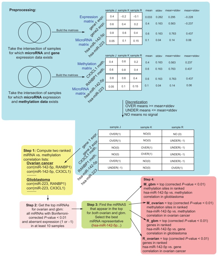
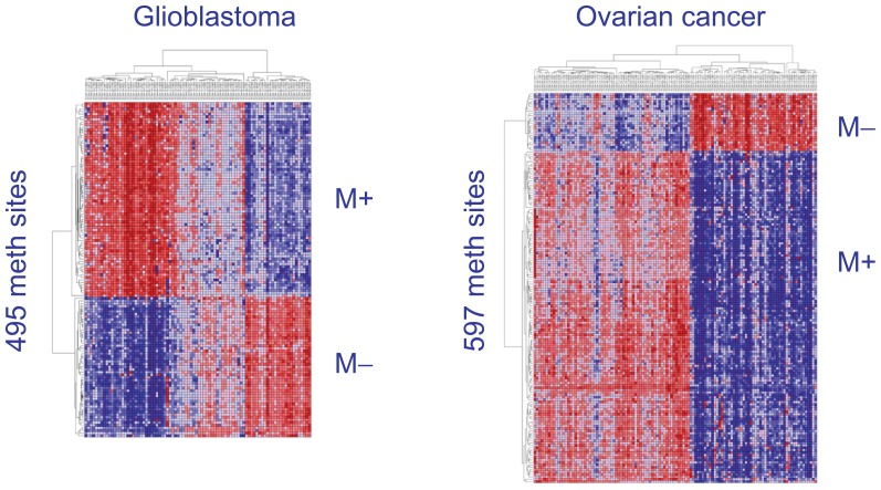
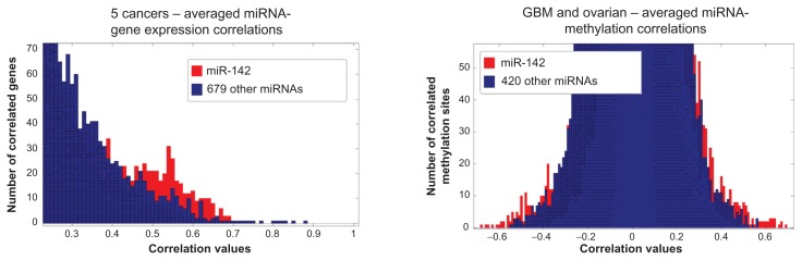
Gene expression profiling has provided insights into different cancer types and revealed tissue-specific expression signatures. Alterations in microRNA expression contribute to the pathogenesis of many types of human diseases. Few studies have integrated all levels of gene expression, miRNA and methylation to uncover correlations between these data types. We performed an integrated profiling to discover instances of miRNAs associated with a gene expression and DNA methylation signature across multiple cancer types. Using data from The Cancer Genome Atlas (TCGA), we revealed a concordant gene expression and methylation signature associated with the microRNA hsa-miR-142 across the same samples. In all cancer types examined, we found a signature of co-expression of a gene set R and methylated sites M, which correlate positively (M+) or negatively (M-) with the expression of hsa-miR-142. The set R consistently contains many genes, such as TRAF3IP3, NCKAP1L, CD53, LAPTM5, PTPRC, EVI2B, DOCK2, LCP2, CYBB and FYB. The signature is preserved across glioblastoma, ovarian, breast, colon, kidney, lung, uterine and rectum cancer. There is 28% overlap of methylation sites in M between glioblastoma (GBM) and ovarian cancer. There is 60% overlap of genes in R between GBM and ovarian (P = 1.3e(-11)). Most of the genes in R are known to be expressed in lymphocytes and haematopoietic stem cells, while M reflects membrane proteins involved in cell-cell adhesion functions. We speculate that the hsa-miR-142 associated signature may signal haematopoietic-specific processes and an accumulation of methylation events triggering a progressive loss of cell-cell adhesion. We also observed that GBM samples belonging to the proneural subtype tend to have underexpressed hsa-miR-142 and R genes, hypomethylated M+ and hypermethylated M-, while the mesenchymal samples have the opposite profile.
基因表达谱分析为不同癌症类型提供了深入见解,并揭示了组织特异性表达特征。微小RNA(miRNA)表达的改变促成了多种人类疾病的发病机制。很少有研究整合基因表达、miRNA和甲基化的所有水平来揭示这些数据类型之间的相关性。我们进行了一项综合分析,以发现跨多种癌症类型与基因表达和DNA甲基化特征相关的miRNA实例。利用来自癌症基因组图谱(TCGA)的数据,我们在相同样本中揭示了与miRNA hsa-miR-142相关的一致基因表达和甲基化特征。在所有检测的癌症类型中,我们发现了一组基因R和甲基化位点M的共表达特征,它们与hsa-miR-142的表达呈正相关(M+)或负相关(M-)。基因集R始终包含许多基因,如TRAF3IP3、NCKAP1L、CD53、LAPTM5、PTPRC、EVI2B、DOCK2、LCP2、CYBB和FYB。该特征在胶质母细胞瘤、卵巢癌、乳腺癌、结肠癌、肾癌、肺癌、子宫癌和直肠癌中均得以保留。胶质母细胞瘤(GBM)和卵巢癌之间甲基化位点M的重叠率为28%。GBM和卵巢癌之间基因集R中的基因重叠率为60%(P = 1.3e(-11))。已知基因集R中的大多数基因在淋巴细胞和造血干细胞中表达,而M反映参与细胞间粘附功能的膜蛋白。我们推测,与hsa-miR-142相关的特征可能标志着造血特异性过程以及甲基化事件的积累,从而引发细胞间粘附的逐渐丧失。我们还观察到,属于神经干细胞亚型的GBM样本往往具有低表达的hsa-miR-142和R基因、低甲基化的M+和高甲基化的M-,而间充质样本则呈现相反的特征。