Integrated Biology of the Gastrointestinal Tract, Institute of Food Research, Norwich, United Kingdom.
PLoS One. 2012;7(5):e37115. doi: 10.1371/journal.pone.0037115. Epub 2012 May 18.
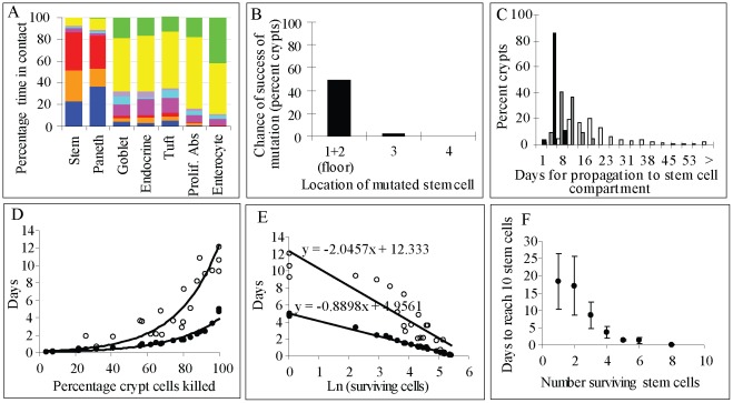
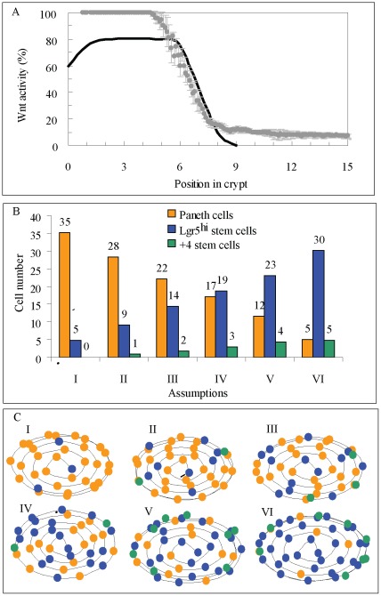
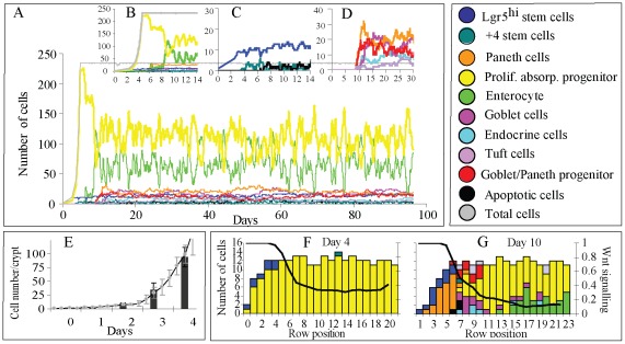
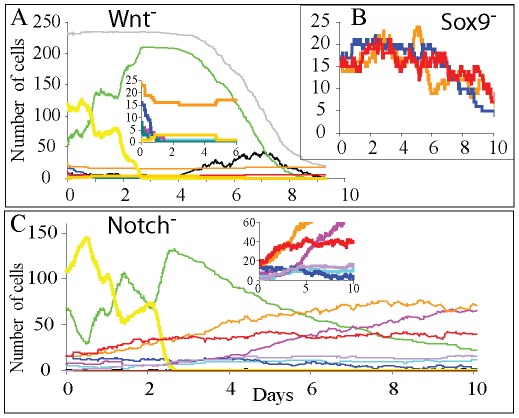
We developed a slow structural relaxation model to describe cellular dynamics in the crypt of the mouse small intestine. Cells are arranged in a three dimensional spiral the size of which dynamically changes according to cell production demands of adjacent villi. Cell differentiation and proliferation is regulated through Wnt and Notch signals, the strength of which depends on the local cell composition. The highest level of Wnt activity is associated with maintaining equipotent stem cells (SC), Paneth cells and common goblet-Paneth cell progenitors (CGPCPs) intermingling at the crypt bottom. Low levels of Wnt signalling area are associated with stem cells giving rise to secretory cells (CGPCPs, enteroendocrine or Tuft cells) and proliferative absorptive progenitors. Deciding between these two fates, secretory and stem/absorptive cells, depends on Notch signalling. Our model predicts that Notch signalling inhibits secretory fate if more than 50% of cells they are in contact with belong to the secretory lineage. CGPCPs under high Wnt signalling will differentiate into Paneth cells while those migrating out from the crypt bottom differentiate into goblet cells. We have assumed that mature Paneth cells migrating upwards undergo anoikis. Structural relaxation explains the localisation of Paneth cells to the crypt bottom in the absence of active forces. The predicted crypt generation time from one SC is 4-5 days with 10-12 days needed to reach a structural steady state. Our predictions are consistent with experimental observations made under altered Wnt and Notch signalling. Mutations affecting stem cells located at the crypt floor have a 50% chance of being propagated throughout the crypt while mutations in cells above are rarely propagated. The predicted recovery time of an injured crypt losing half of its cells is approximately 2 days.
我们开发了一个缓慢的结构弛豫模型来描述小鼠小肠隐窝中的细胞动力学。细胞排列在一个三维螺旋中,其大小根据相邻绒毛的细胞产生需求动态变化。细胞分化和增殖受到 Wnt 和 Notch 信号的调节,其强度取决于局部细胞组成。Wnt 活性的最高水平与维持等效的干细胞(SC)、潘氏细胞和常见的杯状潘氏细胞祖细胞(CGPCP)在隐窝底部混合有关。低水平的 Wnt 信号与干细胞产生分泌细胞(CGPCP、肠内分泌细胞或 Tuft 细胞)和增殖吸收祖细胞有关。在这两种命运(分泌和干细胞/吸收细胞)之间做出决定取决于 Notch 信号。我们的模型预测,如果与它们接触的细胞中有超过 50%属于分泌谱系,Notch 信号将抑制分泌命运。在高 Wnt 信号下,CGPCP 将分化为潘氏细胞,而从隐窝底部迁移出来的细胞则分化为杯状细胞。我们假设向上迁移的成熟潘氏细胞会发生凋亡。结构弛豫解释了在没有主动力的情况下潘氏细胞定位在隐窝底部的原因。从一个 SC 产生隐窝的预测时间为 4-5 天,达到结构稳态需要 10-12 天。我们的预测与改变的 Wnt 和 Notch 信号下的实验观察结果一致。影响位于隐窝底部的干细胞的突变有 50%的机会在整个隐窝中传播,而位于细胞上方的突变则很少传播。失去一半细胞的受损隐窝的恢复时间约为 2 天。