• 文献检索
  • 文档翻译
  • 深度研究
  • 学术资讯
  • Suppr Zotero 插件Zotero 插件
  • 邀请有礼
  • 套餐&价格
  • 历史记录
应用&插件
Suppr Zotero 插件Zotero 插件浏览器插件Mac 客户端Windows 客户端微信小程序
定价
高级版会员购买积分包购买API积分包
服务
文献检索文档翻译深度研究API 文档MCP 服务
关于我们
关于 Suppr公司介绍联系我们用户协议隐私条款
关注我们

Suppr 超能文献

核心技术专利:CN118964589B侵权必究
粤ICP备2023148730 号-1Suppr @ 2026

文献检索

告别复杂PubMed语法,用中文像聊天一样搜索,搜遍4000万医学文献。AI智能推荐,让科研检索更轻松。

立即免费搜索

文件翻译

保留排版,准确专业,支持PDF/Word/PPT等文件格式,支持 12+语言互译。

免费翻译文档

深度研究

AI帮你快速写综述,25分钟生成高质量综述,智能提取关键信息,辅助科研写作。

立即免费体验

结合个体特异性头部模型的高密度扩散光学断层扫描图像质量分析

Image quality analysis of high-density diffuse optical tomography incorporating a subject-specific head model.

作者信息

Zhan Yuxuan, Eggebrecht Adam T, Culver Joseph P, Dehghani Hamid

机构信息

School of Computer Science, University of Birmingham Birmingham, UK.

出版信息

Front Neuroenergetics. 2012 May 24;4:6. doi: 10.3389/fnene.2012.00006. eCollection 2012.

DOI:10.3389/fnene.2012.00006
PMID:22654754
原文链接:https://pmc.ncbi.nlm.nih.gov/articles/PMC3359425/
Abstract

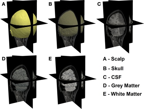
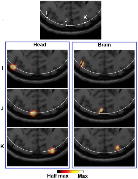
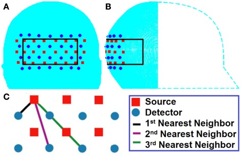
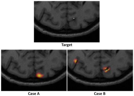
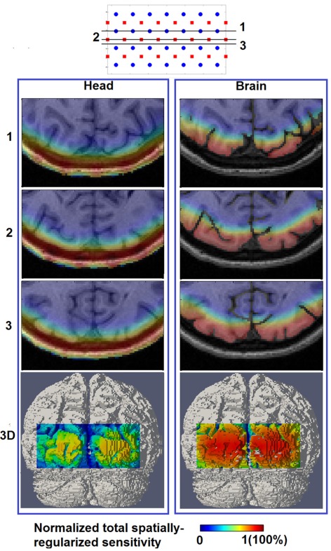
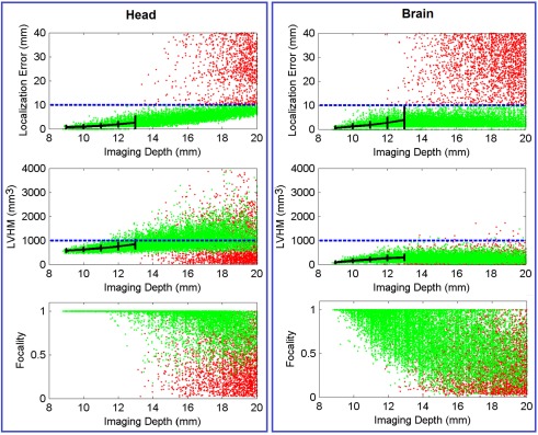
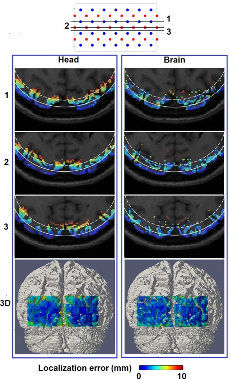
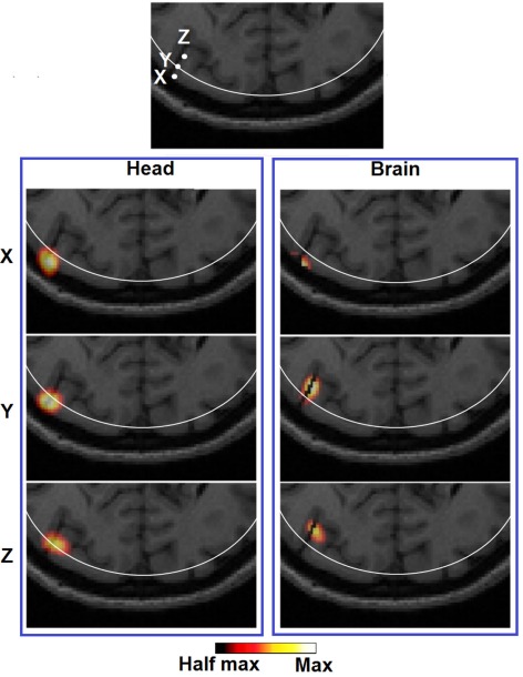
High-density diffuse optical tomography (HD-DOT) methods have shown significant improvement in localization accuracy and image resolution compared to traditional topographic near infrared spectroscopy of the human brain. In this work we provide a comprehensive evaluation of image quality in visual cortex mapping via a simulation study with the use of an anatomical head model derived from MRI data of a human subject. A model of individual head anatomy provides the surface shape and internal structure that allow for the construction of a more realistic physical model for the forward problem, as well as the use of a structural constraint in the inverse problem. The HD-DOT model utilized here incorporates multiple source-detector separations with continuous-wave data with added noise based on experimental results. To evaluate image quality we quantify the localization error and localized volume at half maximum (LVHM) throughout a region of interest within the visual cortex and systematically analyze the use of whole-brain tissue spatial constraint within image reconstruction. Our results demonstrate that an image quality with less than 10 mm in localization error and 1000 m(3) in LVHM can be obtained up to 13 mm below the scalp surface with a typical unconstrained reconstruction and up to 18 mm deep when a whole-brain spatial constraint based on the brain tissue is utilized.

摘要

与传统的人脑近红外光谱地形图相比,高密度漫射光学断层扫描(HD-DOT)方法在定位精度和图像分辨率方面有显著提高。在这项工作中,我们通过使用从人类受试者的MRI数据导出的解剖头部模型进行模拟研究,对视觉皮层映射中的图像质量进行了全面评估。个体头部解剖模型提供了表面形状和内部结构,这有助于构建更逼真的正向问题物理模型,并在逆问题中使用结构约束。这里使用的HD-DOT模型结合了多个源-探测器间距,并根据实验结果对连续波数据添加噪声。为了评估图像质量,我们在视觉皮层的感兴趣区域内量化定位误差和半高宽处的定位体积(LVHM),并系统地分析图像重建中全脑组织空间约束的使用。我们的结果表明,在典型的无约束重建中,在头皮表面以下13毫米处可获得定位误差小于10毫米且LVHM小于1000立方米的图像质量,而在使用基于脑组织的全脑空间约束时,深度可达18毫米。

https://cdn.ncbi.nlm.nih.gov/pmc/blobs/e5a0/3359425/fb4b34ccfa53/fnene-04-00006-g009.jpg
https://cdn.ncbi.nlm.nih.gov/pmc/blobs/e5a0/3359425/18ecf7fc5f39/fnene-04-00006-g001.jpg
https://cdn.ncbi.nlm.nih.gov/pmc/blobs/e5a0/3359425/a5a843200c73/fnene-04-00006-g002.jpg
https://cdn.ncbi.nlm.nih.gov/pmc/blobs/e5a0/3359425/e89e252fc1f1/fnene-04-00006-g003.jpg
https://cdn.ncbi.nlm.nih.gov/pmc/blobs/e5a0/3359425/bef1f1f52081/fnene-04-00006-g004.jpg
https://cdn.ncbi.nlm.nih.gov/pmc/blobs/e5a0/3359425/f3654bc5ee39/fnene-04-00006-g005.jpg
https://cdn.ncbi.nlm.nih.gov/pmc/blobs/e5a0/3359425/3aa333d4e00e/fnene-04-00006-g006.jpg
https://cdn.ncbi.nlm.nih.gov/pmc/blobs/e5a0/3359425/3e52048b0c1a/fnene-04-00006-g007.jpg
https://cdn.ncbi.nlm.nih.gov/pmc/blobs/e5a0/3359425/6e1951bc3da4/fnene-04-00006-g008.jpg
https://cdn.ncbi.nlm.nih.gov/pmc/blobs/e5a0/3359425/fb4b34ccfa53/fnene-04-00006-g009.jpg

相似文献

1
Image quality analysis of high-density diffuse optical tomography incorporating a subject-specific head model.结合个体特异性头部模型的高密度扩散光学断层扫描图像质量分析
Front Neuroenergetics. 2012 May 24;4:6. doi: 10.3389/fnene.2012.00006. eCollection 2012.
2
Atlas-based head modeling and spatial normalization for high-density diffuse optical tomography: in vivo validation against fMRI.基于图谱的头部建模和高密度扩散光学断层成像的空间标准化:与 fMRI 的体内验证。
Neuroimage. 2014 Jan 15;85 Pt 1(0 1):117-26. doi: 10.1016/j.neuroimage.2013.03.069. Epub 2013 Apr 8.
3
Investigation of effect of modulation frequency on high-density diffuse optical tomography image quality.调制频率对高密度扩散光学层析成像图像质量影响的研究
Neurophotonics. 2021 Oct;8(4):045002. doi: 10.1117/1.NPh.8.4.045002. Epub 2021 Nov 24.
4
High-density functional diffuse optical tomography based on frequency-domain measurements improves image quality and spatial resolution.基于频域测量的高密度功能扩散光学断层扫描可提高图像质量和空间分辨率。
Neurophotonics. 2019 Jul;6(3):035007. doi: 10.1117/1.NPh.6.3.035007. Epub 2019 Aug 21.
5
Comparison of source localization techniques in diffuse optical tomography for fNIRS application using a realistic head model.使用逼真头部模型的功能近红外光谱应用中扩散光学层析成像源定位技术的比较
Biomed Opt Express. 2018 Jun 7;9(7):2994-3016. doi: 10.1364/BOE.9.002994. eCollection 2018 Jul 1.
6
A quantitative spatial comparison of high-density diffuse optical tomography and fMRI cortical mapping.高分辨率扩散光学断层成像和 fMRI 皮质映射的定量空间比较。
Neuroimage. 2012 Jul 16;61(4):1120-8. doi: 10.1016/j.neuroimage.2012.01.124. Epub 2012 Feb 10.
7
Simulation study of magnetic resonance imaging-guided cortically constrained diffuse optical tomography of human brain function.磁共振成像引导的人脑功能皮质约束扩散光学断层扫描的模拟研究
Appl Opt. 2005 Apr 1;44(10):1957-68. doi: 10.1364/ao.44.001957.
8
Subject-specific information enhances spatial accuracy of high-density diffuse optical tomography.特定受试者信息可提高高密度扩散光学断层扫描的空间准确性。
Front Neuroergon. 2024 Feb 19;5:1283290. doi: 10.3389/fnrgo.2024.1283290. eCollection 2024.
9
Optimizing the regularization for image reconstruction of cerebral diffuse optical tomography.优化脑扩散光学断层扫描图像重建的正则化方法。
J Biomed Opt. 2014 Sep;19(9):96006. doi: 10.1117/1.JBO.19.9.096006.
10
Quantitative evaluation of high-density diffuse optical tomography: in vivo resolution and mapping performance.高密度扩散光学断层成像的定量评估:体内分辨率和映射性能。
J Biomed Opt. 2010 Mar-Apr;15(2):026006. doi: 10.1117/1.3368999.

引用本文的文献

1
Revisiting equivalent optical properties for cerebrospinal fluid to improve diffusion-based modeling accuracy in the brain.重新审视脑脊液的等效光学特性以提高基于扩散的脑模型精度。
Neurophotonics. 2025 Jan;12(1):015009. doi: 10.1117/1.NPh.12.1.015009. Epub 2025 Feb 14.
2
Diffuse optical tomography of the brain: effects of inaccurate baseline optical parameters and refinements using learned post-processing.大脑的扩散光学断层扫描:不准确的基线光学参数的影响以及使用学习后处理的改进
Biomed Opt Express. 2024 Jul 5;15(8):4470-4485. doi: 10.1364/BOE.524245. eCollection 2024 Aug 1.
3
Revisiting equivalent optical properties for cerebrospinal fluid to improve diffusion-based modeling accuracy in the brain.

本文引用的文献

1
A quantitative spatial comparison of high-density diffuse optical tomography and fMRI cortical mapping.高分辨率扩散光学断层成像和 fMRI 皮质映射的定量空间比较。
Neuroimage. 2012 Jul 16;61(4):1120-8. doi: 10.1016/j.neuroimage.2012.01.124. Epub 2012 Feb 10.
2
Resting-state functional connectivity assessed with two diffuse optical tomographic systems.用两种扩散光学断层成像系统评估静息态功能连接。
J Biomed Opt. 2011 Apr;16(4):046006. doi: 10.1117/1.3561687.
3
Visualization of light propagation in visible Chinese human head for functional near-infrared spectroscopy.
重新审视脑脊液的等效光学特性以提高基于扩散的脑模型精度。
bioRxiv. 2024 Dec 9:2024.08.20.608859. doi: 10.1101/2024.08.20.608859.
4
Subject-specific information enhances spatial accuracy of high-density diffuse optical tomography.特定受试者信息可提高高密度扩散光学断层扫描的空间准确性。
Front Neuroergon. 2024 Feb 19;5:1283290. doi: 10.3389/fnrgo.2024.1283290. eCollection 2024.
5
Diffuse optical reconstructions of functional near infrared spectroscopy data using maximum entropy on the mean.利用均值最大熵对功能近红外光谱数据进行漫射光学重建。
Sci Rep. 2022 Feb 10;12(1):2316. doi: 10.1038/s41598-022-06082-1.
6
Processing pipeline for image reconstructed fNIRS analysis using both MRI templates and individual anatomy.使用MRI模板和个体解剖结构进行图像重建功能近红外光谱分析的处理流程。
Neurophotonics. 2021 Apr;8(2):025010. doi: 10.1117/1.NPh.8.2.025010. Epub 2021 Jun 12.
7
Evaluation of a personalized functional near infra-red optical tomography workflow using maximum entropy on the mean.使用均值上的最大熵评估个性化功能近红外光学断层扫描工作流程。
Hum Brain Mapp. 2021 Oct 15;42(15):4823-4843. doi: 10.1002/hbm.25566. Epub 2021 Aug 3.
8
Best practices for fNIRS publications.功能近红外光谱(fNIRS)出版物的最佳实践。
Neurophotonics. 2021 Jan;8(1):012101. doi: 10.1117/1.NPh.8.1.012101. Epub 2021 Jan 7.
9
Sensitivity Laplacian Ratio-Based Optimization of the Projection Selection for Diffuse Optical Tomography.基于灵敏度拉普拉斯比率的扩散光学层析成像投影选择优化
J Med Signals Sens. 2020 Apr 25;10(2):119-124. doi: 10.4103/jmss.JMSS_67_18. eCollection 2020 Apr-Jun.
10
High-density speckle contrast optical tomography of cerebral blood flow response to functional stimuli in the rodent brain.啮齿动物大脑中对功能刺激的脑血流反应的高密度散斑对比光学断层扫描。
Neurophotonics. 2019 Oct;6(4):045001. doi: 10.1117/1.NPh.6.4.045001. Epub 2019 Oct 8.
可见中国人头部内光传播的可视化用于功能近红外光谱学。
J Biomed Opt. 2011 Apr;16(4):045001. doi: 10.1117/1.3567085.
4
High-resolution optical functional mapping of the human somatosensory cortex.人类躯体感觉皮层的高分辨率光学功能图谱
Front Neuroenergetics. 2010 Jun 14;2:12. doi: 10.3389/fnene.2010.00012. eCollection 2010.
5
Quantitative evaluation of high-density diffuse optical tomography: in vivo resolution and mapping performance.高密度扩散光学断层成像的定量评估:体内分辨率和映射性能。
J Biomed Opt. 2010 Mar-Apr;15(2):026006. doi: 10.1117/1.3368999.
6
Near infrared optical tomography using NIRFAST: Algorithm for numerical model and image reconstruction.使用NIRFAST的近红外光学断层扫描:数值模型与图像重建算法
Commun Numer Methods Eng. 2008 Aug 15;25(6):711-732. doi: 10.1002/cnm.1162.
7
Probabilistic atlas can improve reconstruction from optical imaging of the neonatal brain.概率图谱可以改善新生儿脑部光学成像的重建。
Opt Express. 2009 Aug 17;17(17):14977-92. doi: 10.1364/oe.17.014977.
8
Anatomical atlas-guided diffuse optical tomography of brain activation.基于解剖图谱的脑激活弥散光学层析成像。
Neuroimage. 2010 Jan 1;49(1):561-7. doi: 10.1016/j.neuroimage.2009.07.033. Epub 2009 Jul 28.
9
Phase-encoded retinotopy as an evaluation of diffuse optical neuroimaging.相位编码视网膜地形图作为扩散光学神经影像学的评估方法。
Neuroimage. 2010 Jan 1;49(1):568-77. doi: 10.1016/j.neuroimage.2009.07.023. Epub 2009 Jul 23.
10
Numerical modelling and image reconstruction in diffuse optical tomography.扩散光学层析成像中的数值建模与图像重建
Philos Trans A Math Phys Eng Sci. 2009 Aug 13;367(1900):3073-93. doi: 10.1098/rsta.2009.0090.