Mozumder Meghdoot, Hirvi Pauliina, Nissilä Ilkka, Hauptmann Andreas, Ripoll Jorge, Singh David E
Department of Technical Physics, University of Eastern Finland, P.O. Box 1627, 70211 Kuopio, Finland.
Department of Mathematics and Systems Analysis, Aalto University, P.O. Box 11100, 00076 Aalto, Finland.
Biomed Opt Express. 2024 Jul 5;15(8):4470-4485. doi: 10.1364/BOE.524245. eCollection 2024 Aug 1.
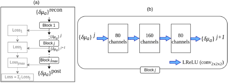
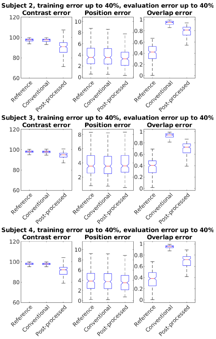
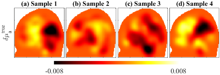
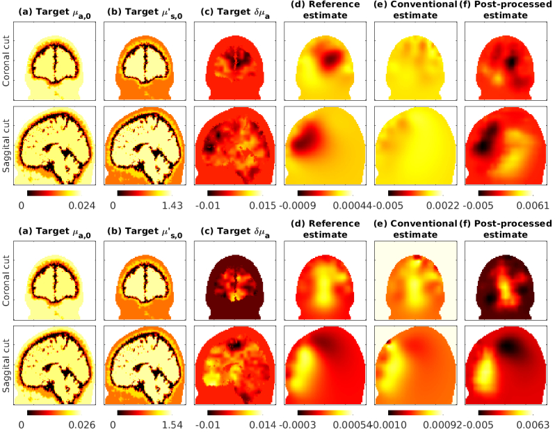
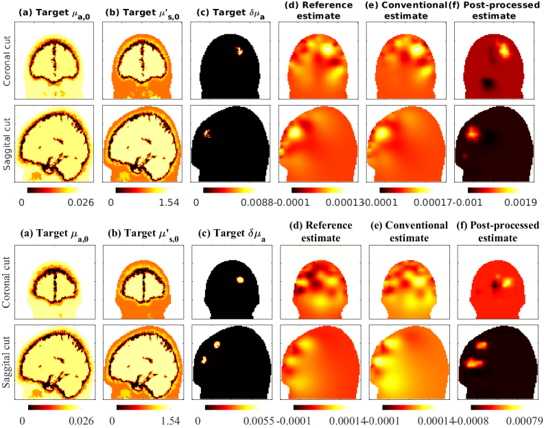
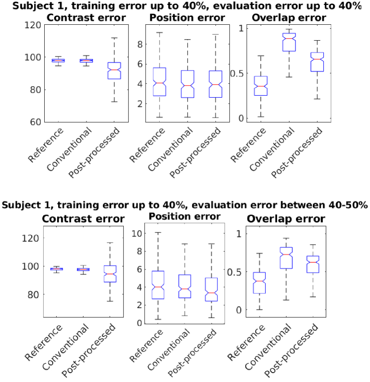
Diffuse optical tomography (DOT) uses near-infrared light to image spatially varying optical parameters in biological tissues. In functional brain imaging, DOT uses a perturbation model to estimate the changes in optical parameters, corresponding to changes in measured data due to brain activity. The perturbation model typically uses approximate baseline optical parameters of the different brain compartments, since the actual baseline optical parameters are unknown. We simulated the effects of these approximate baseline optical parameters using parameter variations earlier reported in literature, and brain atlases from four adult subjects. We report the errors in estimated activation contrast, localization, and area when incorrect baseline values were used. Further, we developed a post-processing technique based on deep learning methods that can reduce the effects due to inaccurate baseline optical parameters. The method improved imaging of brain activation changes in the presence of such errors.
扩散光学层析成像(DOT)利用近红外光对生物组织中空间变化的光学参数进行成像。在功能性脑成像中,DOT使用微扰模型来估计光学参数的变化,这些变化对应于由于大脑活动而导致的测量数据的变化。微扰模型通常使用不同脑区的近似基线光学参数,因为实际的基线光学参数是未知的。我们利用文献中先前报道的参数变化以及来自四名成年受试者的脑图谱,模拟了这些近似基线光学参数的影响。我们报告了使用不正确的基线值时估计激活对比度、定位和面积的误差。此外,我们开发了一种基于深度学习方法的后处理技术,该技术可以减少由于不准确的基线光学参数所带来的影响。该方法改善了在存在此类误差情况下大脑激活变化的成像。