McKay Orthopaedic Research Laboratory, Department of Orthopaedic Surgery, Perelman School of Medicine, University of Pennsylvania, Philadelphia, PA 19104, USA.
J Mech Behav Biomed Mater. 2012 Jul;11:92-101. doi: 10.1016/j.jmbbm.2012.03.006. Epub 2012 Mar 24.
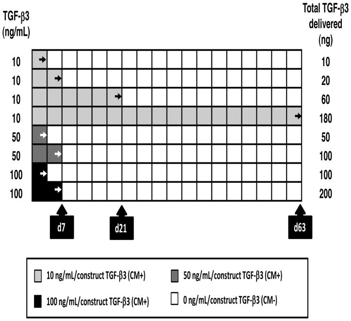
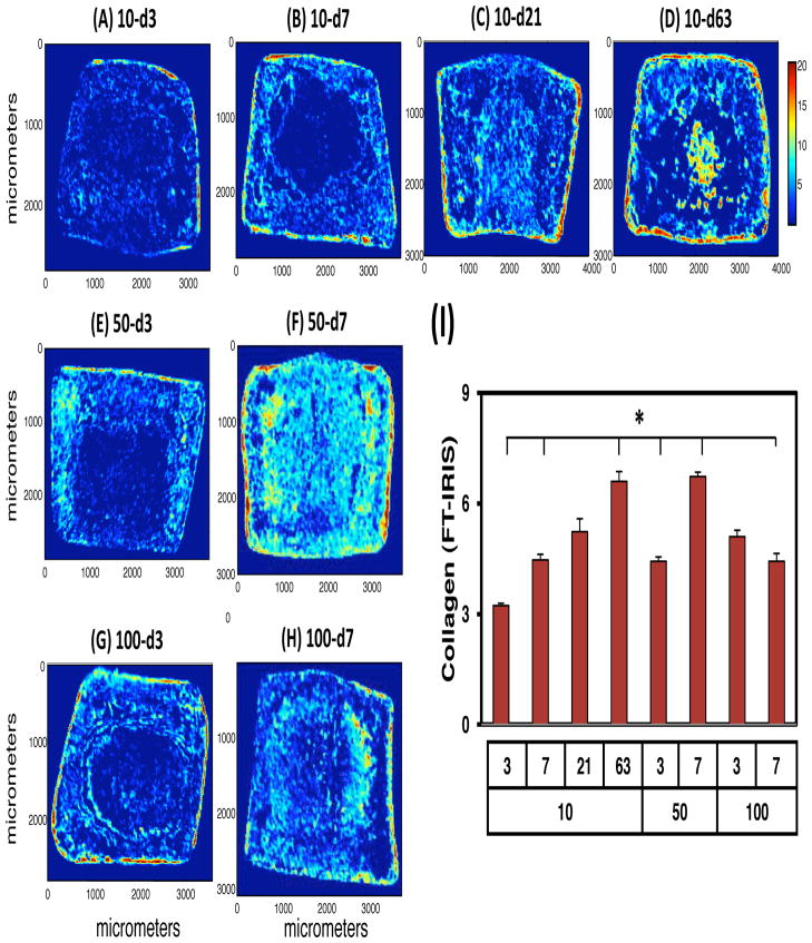
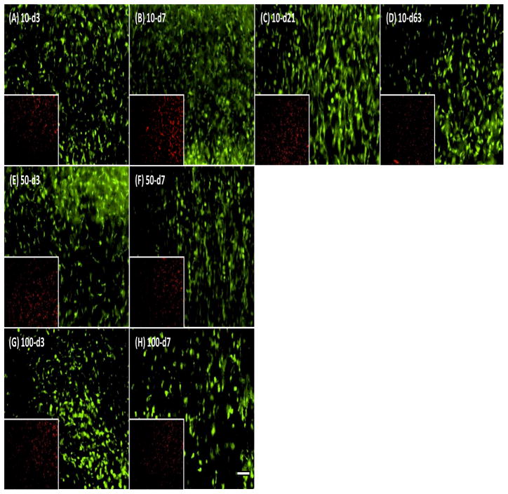
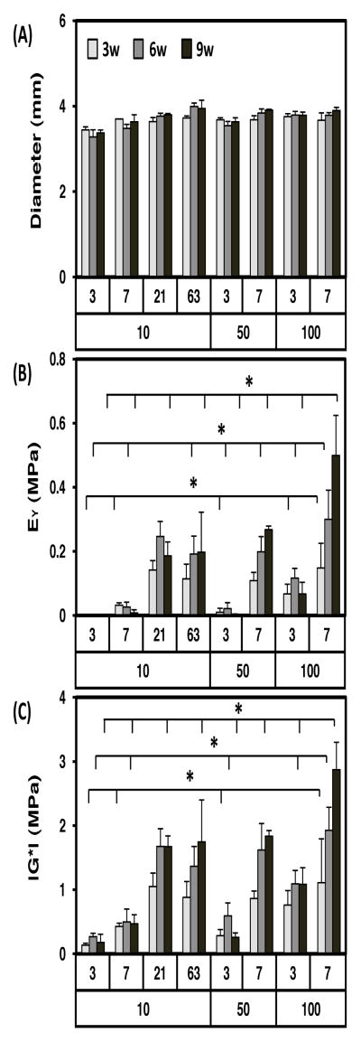
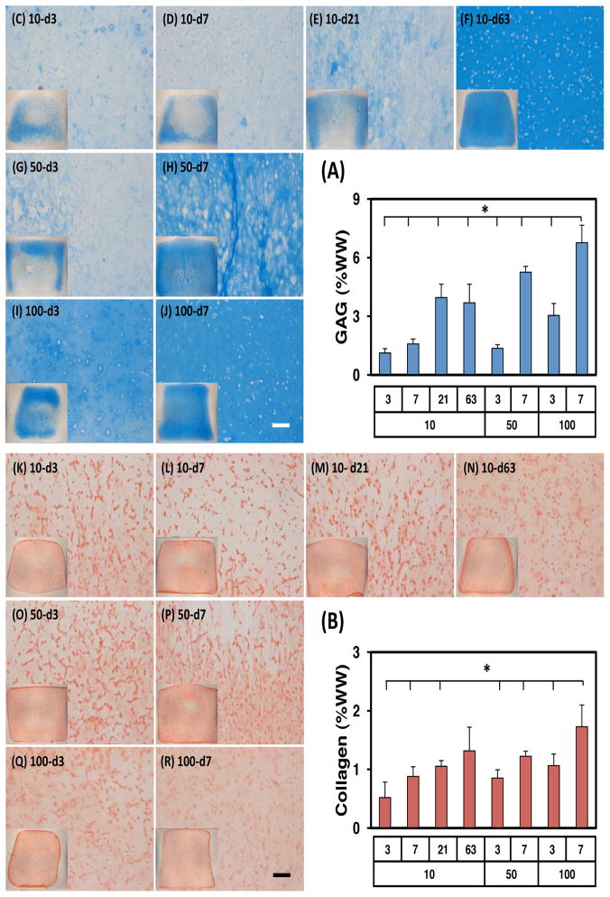
Tissue engineering with adult stem cells is a promising approach for the restoration of focal defects in articular cartilage. For this, progenitor cells would ideally be delivered to (and maintained within) the defect site via a biocompatible material and in combination with soluble factors to promote initial cell differentiation and subsequent tissue maturation in vivo. While growth factor delivery methods are continually being optimized, most offer only a short (days to weeks) delivery profile at high doses. To address this issue, we investigated mesenchymal stem cell (MSC) differentiation and maturation in photocrosslinkable hyaluronic acid (HA) hydrogels with transient exposure to the pro-chondrogenic molecule transforming growth factor-beta3 (TGF-β3), at varying doses (10, 50 and 100 ng/mL) and durations (3, 7, 21 and 63 days). Mechanical, biochemical, and histological outcomes were evaluated through 9 weeks of culture. Results showed that a brief exposure (7 days) to a very high level (100 ng/mL) of TGF-β3 was sufficient to both induce and maintain cartilage formation in these 3D constructs. Indeed, this short delivery resulted in constructs with mechanical and biochemical properties that exceeded that of continuous exposure to a lower level (10 ng/mL) of TGF-β3 over the entire 9-week time course. Of important note, the total TGF delivery in these two scenarios was roughly equivalent (200 vs. 180 ng), but the timing of delivery differed markedly. These data support the idea that acute exposure to a high dose of TGF will induce functional and long-term differentiation of stem cell populations, and further our efforts to improve cartilage repair in vivo.
利用成体干细胞进行组织工程是修复关节软骨局灶性缺损的一种很有前途的方法。为此,理想情况下,祖细胞应通过生物相容性材料递送到(并保持在)缺陷部位,并与可溶性因子结合,以促进初始细胞分化,并随后在体内进行组织成熟。虽然生长因子递送方法在不断得到优化,但大多数方法仅能以高剂量提供短期(数天至数周)的递送。为了解决这个问题,我们研究了间充质干细胞(MSC)在可光交联透明质酸(HA)水凝胶中的分化和成熟,该水凝胶短暂暴露于前软骨形成分子转化生长因子-β3(TGF-β3),剂量(10、50 和 100 ng/mL)和时间(3、7、21 和 63 天)不同。通过 9 周的培养评估了机械、生化和组织学结果。结果表明,短暂暴露(7 天)于非常高水平(100 ng/mL)的 TGF-β3 足以诱导和维持这些 3D 构建体中的软骨形成。事实上,这种短期的递送导致构建体具有的机械和生化特性超过了在整个 9 周时间过程中持续暴露于较低水平(10 ng/mL)的 TGF-β3。值得注意的是,这两种情况下的总 TGF 递送大致相当(200 与 180 ng),但递送时间差异显著。这些数据支持这样一种观点,即急性暴露于高剂量 TGF 将诱导干细胞群体的功能性和长期分化,并进一步推动我们改善体内软骨修复的努力。