• 文献检索
  • 文档翻译
  • 深度研究
  • 学术资讯
  • Suppr Zotero 插件Zotero 插件
  • 邀请有礼
  • 套餐&价格
  • 历史记录
应用&插件
Suppr Zotero 插件Zotero 插件浏览器插件Mac 客户端Windows 客户端微信小程序
定价
高级版会员购买积分包购买API积分包
服务
文献检索文档翻译深度研究API 文档MCP 服务
关于我们
关于 Suppr公司介绍联系我们用户协议隐私条款
关注我们

Suppr 超能文献

核心技术专利:CN118964589B侵权必究
粤ICP备2023148730 号-1Suppr @ 2026

文献检索

告别复杂PubMed语法,用中文像聊天一样搜索,搜遍4000万医学文献。AI智能推荐,让科研检索更轻松。

立即免费搜索

文件翻译

保留排版,准确专业,支持PDF/Word/PPT等文件格式,支持 12+语言互译。

免费翻译文档

深度研究

AI帮你快速写综述,25分钟生成高质量综述,智能提取关键信息,辅助科研写作。

立即免费体验

肺部 MRI(1/3):方法。

MRI of the lung (1/3): methods.

机构信息

Academic Radiology, Royal Hallamshire Hospital Sheffield, University of Sheffield, Sheffield, S10 2JF, UK,

出版信息

Insights Imaging. 2012 Aug;3(4):345-53. doi: 10.1007/s13244-012-0176-x. Epub 2012 Jun 13.

DOI:10.1007/s13244-012-0176-x
PMID:22695952
原文链接:https://pmc.ncbi.nlm.nih.gov/articles/PMC3481083/
Abstract

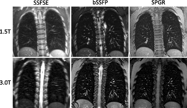
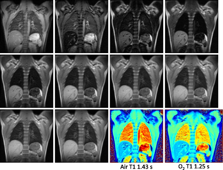
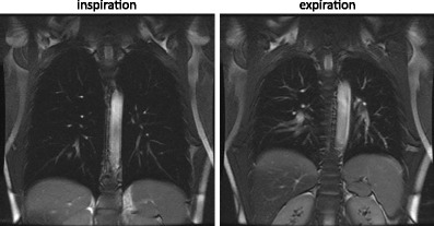
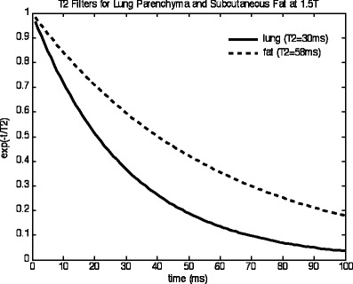
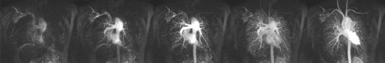
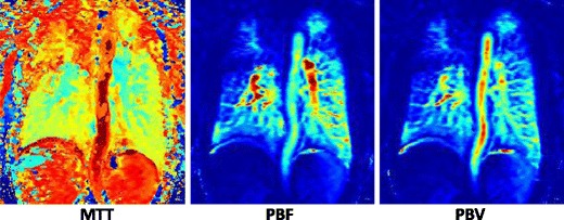
Proton magnetic resonance imaging (MRI) has recently emerged as a clinical tool to image the lungs. This paper outlines the current technical aspects of MRI pulse sequences, radiofrequency (RF) coils and MRI system requirements needed for imaging the pulmonary parenchyma and vasculature. Lung MRI techniques are presented as a "technical toolkit", from which MR protocols will be composed in the subsequent papers for comprehensive imaging of lung disease and function (parts 2 and 3). This paper is pitched at MR scientists, technicians and radiologists who are interested in understanding and establishing lung MRI methods. Images from a 1.5 T scanner are used for illustration of the sequences and methods that are highlighted. Main Messages • Outline of the hardware and pulse sequence requirements for proton lung MRI • Overview of pulse sequences for lung parenchyma, vascular and functional imaging with protons • Demonstration of the pulse-sequence building blocks for clinical lung MRI protocols.

摘要

质子磁共振成像(MRI)最近已成为一种临床工具,可用于肺部成像。本文概述了用于肺部实质和血管成像的 MRI 脉冲序列、射频(RF)线圈和 MRI 系统要求的当前技术方面。肺部 MRI 技术被呈现为一个“技术工具包”,后续的论文将根据这些技术组成用于全面成像肺部疾病和功能的 MR 协议(第 2 部分和第 3 部分)。本文面向对理解和建立肺部 MRI 方法感兴趣的磁共振科学家、技术人员和放射科医生。使用 1.5T 扫描仪的图像来说明突出显示的序列和方法。主要信息 • 质子肺部 MRI 的硬件和脉冲序列要求概述 • 用于肺部实质、血管和功能成像的脉冲序列概述 • 临床肺部 MRI 协议中脉冲序列构建模块的演示

https://cdn.ncbi.nlm.nih.gov/pmc/blobs/9a34/3481083/268f3a3f7d54/13244_2012_176_Fig10_HTML.jpg
https://cdn.ncbi.nlm.nih.gov/pmc/blobs/9a34/3481083/09211762fa84/13244_2012_176_Fig1_HTML.jpg
https://cdn.ncbi.nlm.nih.gov/pmc/blobs/9a34/3481083/6880c31dd7c5/13244_2012_176_Fig2_HTML.jpg
https://cdn.ncbi.nlm.nih.gov/pmc/blobs/9a34/3481083/659d768bcf79/13244_2012_176_Fig3_HTML.jpg
https://cdn.ncbi.nlm.nih.gov/pmc/blobs/9a34/3481083/734070aa1ada/13244_2012_176_Fig4_HTML.jpg
https://cdn.ncbi.nlm.nih.gov/pmc/blobs/9a34/3481083/28b9645a5aa5/13244_2012_176_Fig5_HTML.jpg
https://cdn.ncbi.nlm.nih.gov/pmc/blobs/9a34/3481083/1c8b7bb49d53/13244_2012_176_Fig6_HTML.jpg
https://cdn.ncbi.nlm.nih.gov/pmc/blobs/9a34/3481083/c679c9b833f6/13244_2012_176_Fig7_HTML.jpg
https://cdn.ncbi.nlm.nih.gov/pmc/blobs/9a34/3481083/5c52132bb8be/13244_2012_176_Fig8_HTML.jpg
https://cdn.ncbi.nlm.nih.gov/pmc/blobs/9a34/3481083/b8de40ee4534/13244_2012_176_Fig9_HTML.jpg
https://cdn.ncbi.nlm.nih.gov/pmc/blobs/9a34/3481083/268f3a3f7d54/13244_2012_176_Fig10_HTML.jpg

相似文献

1
MRI of the lung (1/3): methods.肺部 MRI(1/3):方法。
Insights Imaging. 2012 Aug;3(4):345-53. doi: 10.1007/s13244-012-0176-x. Epub 2012 Jun 13.
2
Review of oxygen-enhanced lung mri: Pulse sequences for image acquisition and T measurement.氧增强肺部MRI综述:用于图像采集和T测量的脉冲序列
Med Phys. 2023 Oct;50(10):5987-6007. doi: 10.1002/mp.16553. Epub 2023 Jun 21.
3
Canadian Society of Thoracic Radiology/Canadian Association of Radiologists Clinical Practice Guidance for Non-Vascular Thoracic MRI.加拿大胸放射学会/加拿大放射学家协会非血管性胸部MRI临床实践指南
Can Assoc Radiol J. 2021 Nov;72(4):831-845. doi: 10.1177/0846537121998961. Epub 2021 Mar 29.
4
Quantitative analysis of image quality for acceptance and commissioning of an MRI simulator with a semiautomatic method.采用半自动方法对MRI模拟机验收和调试的图像质量进行定量分析。
J Appl Clin Med Phys. 2018 May;19(3):326-335. doi: 10.1002/acm2.12311. Epub 2018 Mar 24.
5
Building blocks for thoracic MRI: Challenges, sequences, and protocol design.胸部 MRI 的构建模块:挑战、序列和方案设计。
J Magn Reson Imaging. 2019 Sep;50(3):682-701. doi: 10.1002/jmri.26677. Epub 2019 Feb 19.
6
Rapid and robust pulmonary proton ZTE imaging in the mouse.小鼠肺部质子 ZTE 的快速且稳健成像
NMR Biomed. 2014 Sep;27(9):1129-34. doi: 10.1002/nbm.3161. Epub 2014 Jul 26.
7
Proton MRI of the Lung: How to Tame Scarce Protons and Fast Signal Decay.肺部质子 MRI:如何驯服稀缺的质子和快速衰减的信号。
J Magn Reson Imaging. 2021 May;53(5):1344-1357. doi: 10.1002/jmri.27122. Epub 2020 Mar 12.
8
Simultaneous PET/MR imaging: MR-based attenuation correction of local radiofrequency surface coils.同时进行的正电子发射断层扫描/磁共振成像:基于磁共振的局部射频表面线圈衰减校正。
Med Phys. 2012 Jul;39(7):4306-15. doi: 10.1118/1.4729716.
9
Magnetic resonance imaging of the genitourinary tract.泌尿生殖道的磁共振成像
Urol Clin North Am. 1986 Aug;13(3):531-50.
10
Lung MRI at 1.5 and 3 Tesla: observer preference study and lesion contrast using five different pulse sequences.1.5和3特斯拉的肺部磁共振成像:观察者偏好研究及使用五种不同脉冲序列的病变对比度
Invest Radiol. 2007 Jun;42(6):377-83. doi: 10.1097/01.rli.0000261926.86278.96.

引用本文的文献

1
Plug-and-Play Self-Supervised Denoising for Pulmonary Perfusion MRI.用于肺部灌注MRI的即插即用自监督去噪
Bioengineering (Basel). 2025 Jul 1;12(7):724. doi: 10.3390/bioengineering12070724.
2
[Magnetic resonance imaging of the pediatric chest].[小儿胸部的磁共振成像]
Radiologie (Heidelb). 2025 Jul 9. doi: 10.1007/s00117-025-01486-2.
3
Zero-echo time magnetic resonance imaging for visualizing pulmonary neoplasms.用于可视化肺部肿瘤的零回波时间磁共振成像

本文引用的文献

1
MRI of the lung (2/3). Why … when … how?肺部磁共振成像(二/三)。为什么……何时……如何?
Insights Imaging. 2012 Aug;3(4):355-71. doi: 10.1007/s13244-011-0146-8. Epub 2012 Feb 13.
2
MRI of the lung (3/3)-current applications and future perspectives.肺部 MRI(3/3)——当前的应用与未来展望。
Insights Imaging. 2012 Aug;3(4):373-86. doi: 10.1007/s13244-011-0142-z. Epub 2012 Jan 15.
3
Diagnostic accuracy of contrast-enhanced MR angiography and unenhanced proton MR imaging compared with CT pulmonary angiography in chronic thromboembolic pulmonary hypertension.
BMC Med Imaging. 2025 Jul 7;25(1):274. doi: 10.1186/s12880-025-01666-5.
4
Longitudinal relaxation rate ( ) in lung: a systematic review and meta-analysis.肺的纵向弛豫率( ):一项系统评价和荟萃分析。
MAGMA. 2025 May 16. doi: 10.1007/s10334-025-01259-x.
5
Unlocking the Potential of Chest MRI: Strategies for Establishing a Successful Practice.挖掘胸部磁共振成像的潜力:建立成功实践的策略。
J Korean Soc Radiol. 2025 Jan;86(1):83-104. doi: 10.3348/jksr.2024.0056. Epub 2025 Jan 20.
6
A synthetic lung model (ASYLUM) for validation of functional lung imaging methods shows significant differences between signal-based and deformation-field-based ventilation measurements.一种用于验证功能性肺成像方法的合成肺模型(ASYLUM)显示,基于信号的通气测量和基于变形场的通气测量之间存在显著差异。
Front Med (Lausanne). 2024 Sep 4;11:1418052. doi: 10.3389/fmed.2024.1418052. eCollection 2024.
7
Imaging the Lung in ARDS: A Primer.急性呼吸窘迫综合征肺部影像学:基础篇。
Respir Care. 2024 Jul 24;69(8):1011-1024. doi: 10.4187/respcare.12061.
8
Free-Breathing Functional Pulmonary Proton MRI: A Novel Approach Using Voxel-Wise Lung Ventilation (VOLVE) Assessment in Healthy Volunteers and Patients With Chronic Obstructive Pulmonary Disease.自由呼吸功能性肺部质子磁共振成像:一种在健康志愿者和慢性阻塞性肺疾病患者中使用体素级肺通气(VOLVE)评估的新方法。
J Magn Reson Imaging. 2025 Feb;61(2):663-675. doi: 10.1002/jmri.29444. Epub 2024 May 31.
9
Associated radiation exposure from medical imaging and excess lifetime risk of developing cancer in pediatric patients with pulmonary hypertension.医学成像相关辐射暴露与小儿肺动脉高压患者患癌的终生额外风险
Pulm Circ. 2023 Aug 21;13(3):e12282. doi: 10.1002/pul2.12282. eCollection 2023 Jul.
10
Improving pulmonary perfusion assessment by dynamic contrast-enhanced computed tomography in an experimental lung injury model.利用动态对比增强 CT 提高实验性肺损伤模型中的肺灌注评估。
J Appl Physiol (1985). 2023 Jun 1;134(6):1496-1507. doi: 10.1152/japplphysiol.00159.2023. Epub 2023 May 11.
对比增强磁共振血管造影和质子磁共振成像与 CT 肺动脉造影在慢性血栓栓塞性肺动脉高压中的诊断准确性比较。
Eur Radiol. 2012 Feb;22(2):310-7. doi: 10.1007/s00330-011-2252-x. Epub 2011 Sep 2.
4
Magnetic resonance imaging and computed tomography of respiratory mechanics.呼吸力学的磁共振成像和计算机断层扫描。
J Magn Reson Imaging. 2010 Dec;32(6):1388-97. doi: 10.1002/jmri.22386.
5
Half-Fourier-acquisition single-shot turbo spin-echo (HASTE) MRI of the lung at 3 Tesla using parallel imaging with 32-receiver channel technology.在3特斯拉场强下,采用具有32通道接收技术的并行成像的肺部半傅里叶采集单次激发快速自旋回波(HASTE)磁共振成像。
J Magn Reson Imaging. 2009 Sep;30(3):541-6. doi: 10.1002/jmri.21882.
6
Non-contrast-enhanced perfusion and ventilation assessment of the human lung by means of fourier decomposition in proton MRI.通过质子磁共振成像中的傅里叶分解对人体肺部进行非对比增强灌注和通气评估。
Magn Reson Med. 2009 Sep;62(3):656-64. doi: 10.1002/mrm.22031.
7
Lung imaging under free-breathing conditions.自由呼吸条件下的肺部成像。
Magn Reson Med. 2009 Mar;61(3):723-7. doi: 10.1002/mrm.21846.
8
Flow-compensated self-gating.血流补偿自门控
MAGMA. 2008 Sep;21(5):307-15. doi: 10.1007/s10334-008-0131-5. Epub 2008 Jul 31.
9
Oxygen-enhanced magnetic resonance imaging: influence of different gas delivery methods on the T1-changes of the lungs.氧增强磁共振成像:不同气体输送方法对肺T1变化的影响
Invest Radiol. 2008 Jun;43(6):427-32. doi: 10.1097/RLI.0b013e318169012d.
10
In vivo Gd-DTPA concentration for MR lung perfusion measurements: assessment with computed tomography in a porcine model.用于磁共振肺灌注测量的体内钆喷酸葡胺浓度:在猪模型中用计算机断层扫描进行评估
Eur Radiol. 2008 Oct;18(10):2102-7. doi: 10.1007/s00330-008-0974-1. Epub 2008 Apr 30.