Zempel John M, Politte David G, Kelsey Matthew, Verner Ryan, Nolan Tracy S, Babajani-Feremi Abbas, Prior Fred, Larson-Prior Linda J
Department of Neurology, Washington University School of Medicine, St. Louis MO, USA.
Front Neurol. 2012 Jun 12;3:76. doi: 10.3389/fneur.2012.00076. eCollection 2012.
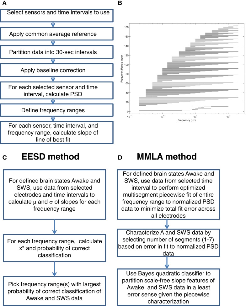
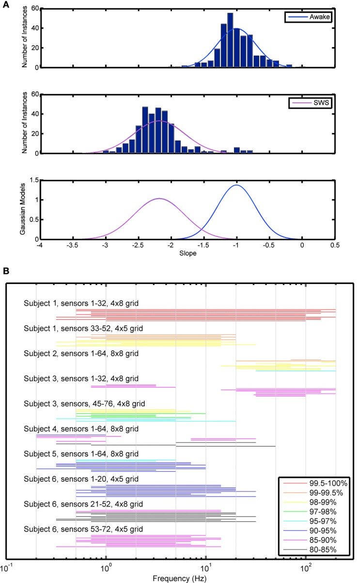
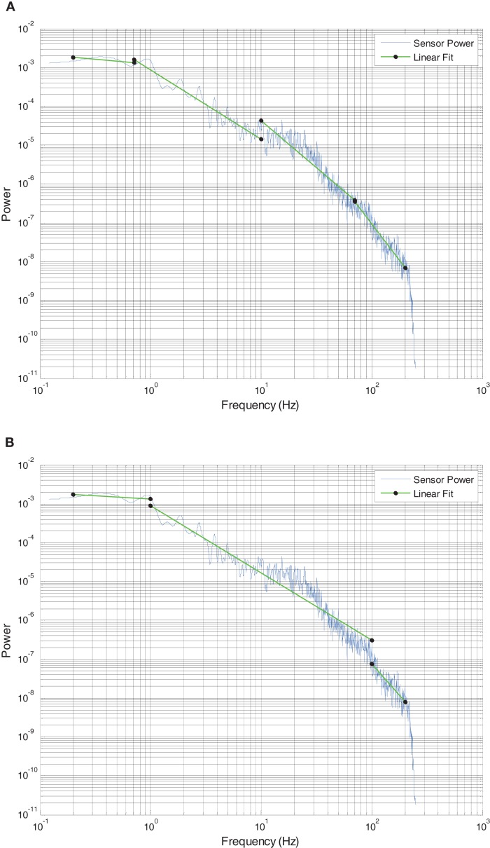
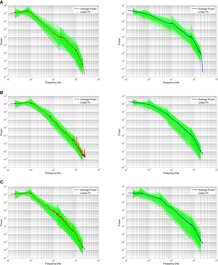
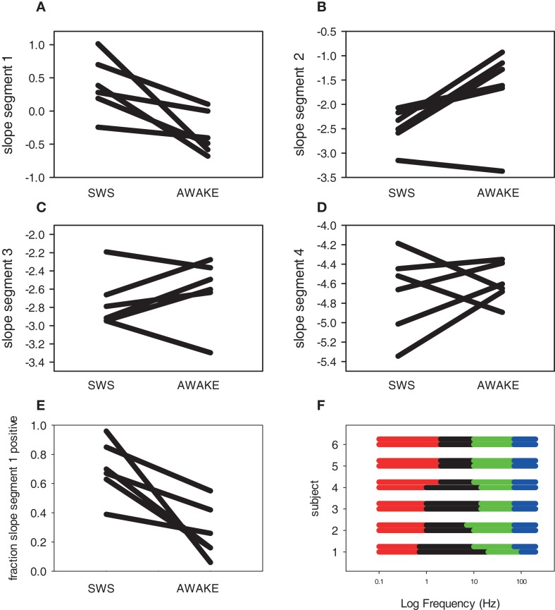
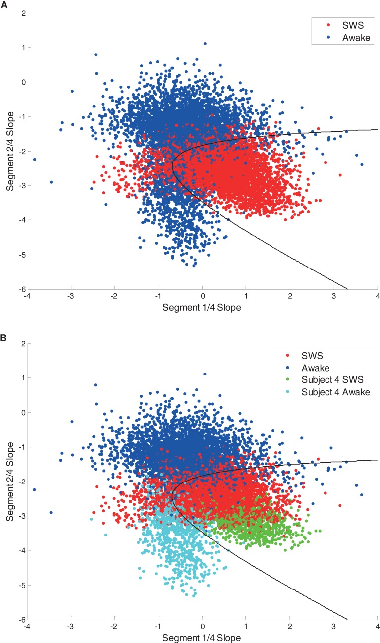
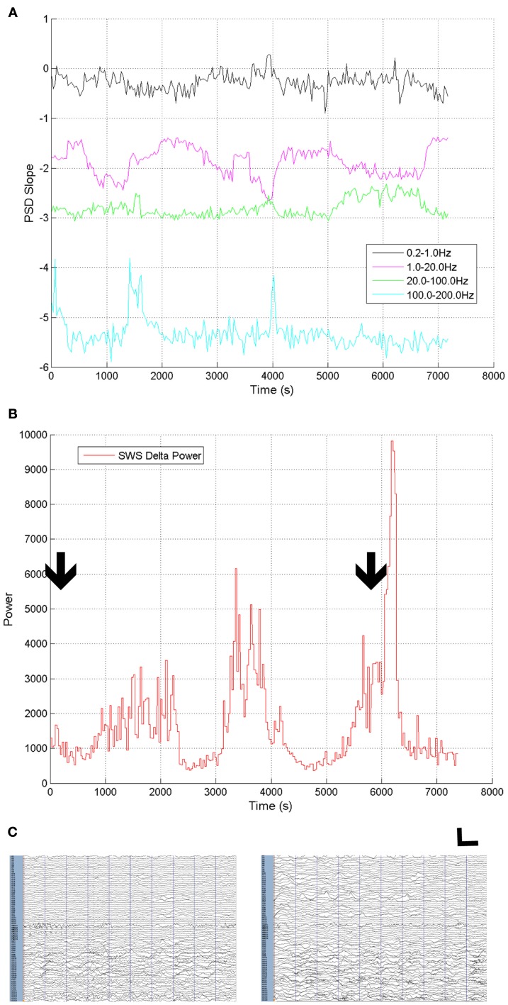
Like many complex dynamic systems, the brain exhibits scale-free dynamics that follow power-law scaling. Broadband power spectral density (PSD) of brain electrical activity exhibits state-dependent power-law scaling with a log frequency exponent that varies across frequency ranges. Widely divergent naturally occurring neural states, awake and slow wave sleep (SWS), were used to evaluate the nature of changes in scale-free indices of brain electrical activity. We demonstrate two analytic approaches to characterizing electrocorticographic (ECoG) data obtained during awake and SWS states. A data-driven approach was used, characterizing all available frequency ranges. Using an equal error state discriminator (EESD), a single frequency range did not best characterize state across data from all six subjects, though the ability to distinguish awake and SWS ECoG data in individual subjects was excellent. Multi-segment piecewise linear fits were used to characterize scale-free slopes across the entire frequency range (0.2-200 Hz). These scale-free slopes differed between awake and SWS states across subjects, particularly at frequencies below 10 Hz and showed little difference at frequencies above 70 Hz. A multivariate maximum likelihood analysis (MMLA) method using the multi-segment slope indices successfully categorized ECoG data in most subjects, though individual variation was seen. In exploring the differences between awake and SWS ECoG data, these analytic techniques show that no change in a single frequency range best characterizes differences between these two divergent biological states. With increasing computational tractability, the use of scale-free slope values to characterize ECoG and EEG data will have practical value in clinical and research studies.
与许多复杂的动态系统一样,大脑呈现出遵循幂律缩放的无标度动态。大脑电活动的宽带功率谱密度(PSD)呈现出与状态相关的幂律缩放,其对数频率指数在不同频率范围内变化。我们利用广泛存在的、截然不同的自然神经状态——清醒和慢波睡眠(SWS),来评估大脑电活动无标度指数变化的本质。我们展示了两种分析方法,用于表征在清醒和SWS状态下获得的皮质脑电图(ECoG)数据。我们采用了一种数据驱动的方法,对所有可用的频率范围进行表征。使用等误差状态鉴别器(EESD)时,单一频率范围并不能最好地表征来自所有六名受试者的数据中的状态,尽管在个体受试者中区分清醒和SWS的ECoG数据的能力非常出色。多段分段线性拟合用于表征整个频率范围(0.2 - 200 Hz)内的无标度斜率。这些无标度斜率在不同受试者的清醒和SWS状态之间存在差异,特别是在低于10 Hz的频率处,而在高于70 Hz的频率处差异不大。使用多段斜率指数的多元最大似然分析(MMLA)方法在大多数受试者中成功地对ECoG数据进行了分类,不过也存在个体差异。在探索清醒和SWS的ECoG数据之间的差异时,这些分析技术表明,单一频率范围内的变化并不能最好地表征这两种不同生物状态之间的差异。随着计算可处理性的提高,使用无标度斜率值来表征ECoG和脑电图数据在临床和研究中具有实际价值。