Kothapalli Sri-Rajasekhar, Liu Hongguang, Liao Joseph C, Cheng Zhen, Gambhir Sanjiv Sam
Biomed Opt Express. 2012 Jun 1;3(6):1215-25. doi: 10.1364/BOE.3.001215. Epub 2012 May 3.
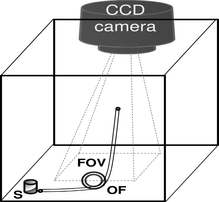
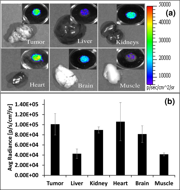
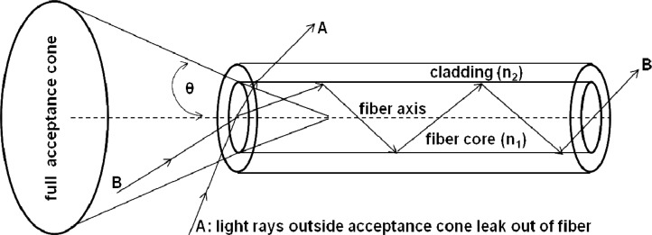
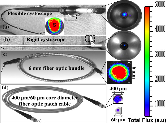
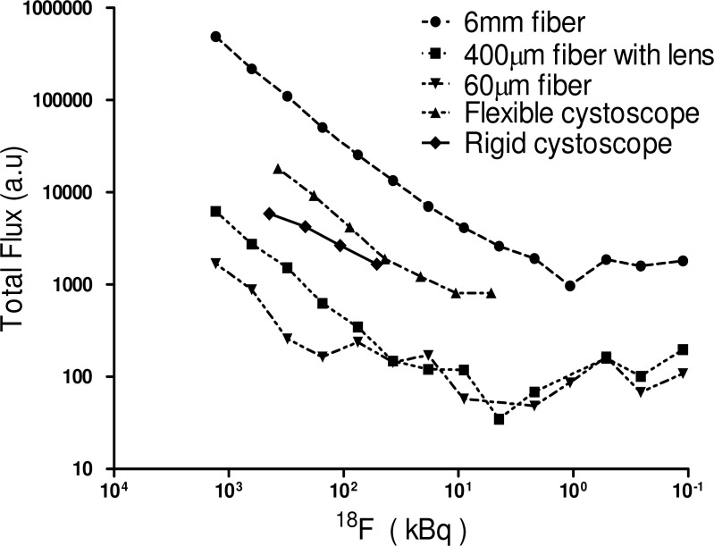
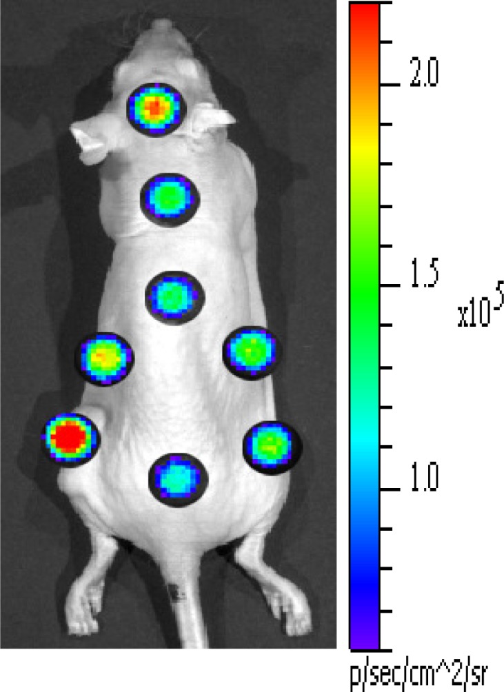
We demonstrate feasibility of endoscopic imaging of Cerenkov light originated when charged nuclear particles, emitted from radionuclides, travel through a biological tissue of living subjects at superluminal velocity. The endoscopy imaging system consists of conventional optical fiber bundle/ clinical endoscopes, an optical imaging lens system, and a sensitive low-noise charge coupled device (CCD) camera. Our systematic studies using phantom samples show that Cerenkov light from as low as 1 µCi of radioactivity emitted from (18)F-Fluorodeoxyglucose (FDG) can be coupled and transmitted through conventional optical fibers and endoscopes. In vivo imaging experiments with tumor bearing mice, intravenously administered with (18)F-FDG, further demonstrated that Cerenkov luminescence endoscopy is a promising new tool in the field of endoscopic molecular imaging.
我们证明了对切伦科夫光进行内镜成像的可行性,这种光源于放射性核素发射的带电核粒子以超光速穿过活体受试者的生物组织时产生。内镜成像系统由传统光纤束/临床内镜、光学成像透镜系统以及灵敏的低噪声电荷耦合器件(CCD)相机组成。我们使用模拟样品进行的系统研究表明,来自低至1微居里(18)F - 氟脱氧葡萄糖(FDG)发射的放射性的切伦科夫光能够通过传统光纤和内镜进行耦合和传输。对静脉注射(18)F - FDG的荷瘤小鼠进行的体内成像实验进一步证明,切伦科夫发光内镜是内镜分子成像领域一种有前景的新工具。