Golan Lior, Yeheskely-Hayon Daniella, Minai Limor, Dann Eldad J, Yelin Dvir
Biomed Opt Express. 2012 Jun 1;3(6):1455-64. doi: 10.1364/BOE.3.001455. Epub 2012 May 21.
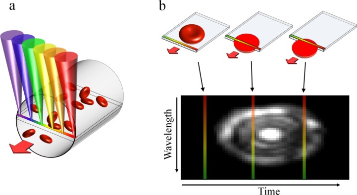
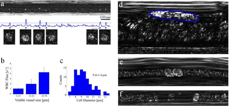
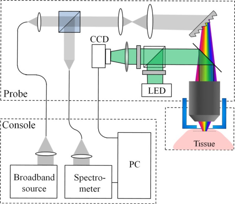
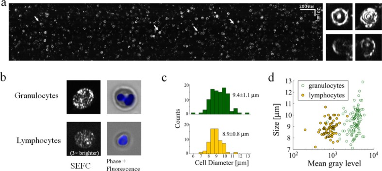
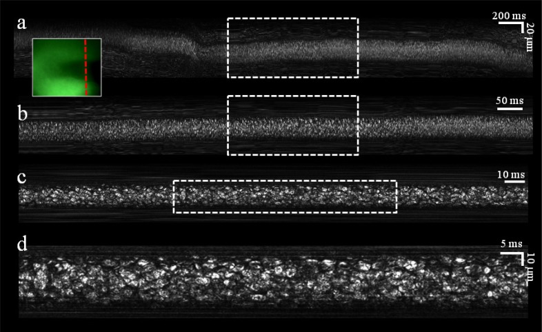
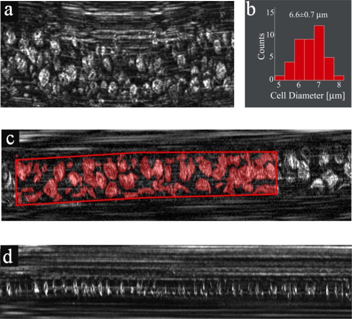
Optical microscopy of blood cells in vivo provides a unique opportunity for clinicians and researchers to visualize the morphology and dynamics of circulating cells, but is usually limited by the imaging speed and by the need for exogenous labeling of the cells. Here we present a label-free approach for in vivo flow cytometry of blood using a compact imaging probe that could be adapted for bedside real-time imaging of patients in clinical settings, and demonstrate subcellular resolution imaging of red and white blood cells flowing in the oral mucosa of a human volunteer. By analyzing the large data sets obtained by the system, valuable blood parameters could be extracted and used for direct, reliable assessment of patient physiology.
体内血细胞的光学显微镜检查为临床医生和研究人员提供了一个独特的机会,使其能够观察循环细胞的形态和动态,但通常受到成像速度和细胞外源性标记需求的限制。在此,我们展示了一种使用紧凑型成像探头进行体内血液流式细胞术的无标记方法,该探头可适用于临床环境中对患者进行床边实时成像,并展示了在人类志愿者口腔黏膜中流动的红细胞和白细胞的亚细胞分辨率成像。通过分析该系统获得的大量数据集,可以提取有价值的血液参数,并用于直接、可靠地评估患者的生理状况。