• 文献检索
  • 文档翻译
  • 深度研究
  • 学术资讯
  • Suppr Zotero 插件Zotero 插件
  • 邀请有礼
  • 套餐&价格
  • 历史记录
应用&插件
Suppr Zotero 插件Zotero 插件浏览器插件Mac 客户端Windows 客户端微信小程序
定价
高级版会员购买积分包购买API积分包
服务
文献检索文档翻译深度研究API 文档MCP 服务
关于我们
关于 Suppr公司介绍联系我们用户协议隐私条款
关注我们

Suppr 超能文献

核心技术专利:CN118964589B侵权必究
粤ICP备2023148730 号-1Suppr @ 2026

文献检索

告别复杂PubMed语法,用中文像聊天一样搜索,搜遍4000万医学文献。AI智能推荐,让科研检索更轻松。

立即免费搜索

文件翻译

保留排版,准确专业,支持PDF/Word/PPT等文件格式,支持 12+语言互译。

免费翻译文档

深度研究

AI帮你快速写综述,25分钟生成高质量综述,智能提取关键信息,辅助科研写作。

立即免费体验

颅内动脉瘤的数字减影血管造影容积三维重建:图文综述。

DSA volumetric 3D reconstructions of intracranial aneurysms: A pictorial essay.

作者信息

Cieściński Jakub, Serafin Zbigniew, Strześniewski Piotr, Lasek Władysław, Beuth Wojciech

机构信息

Department of Radiology and Diagnostic Imaging, Nicolaus Copernicus University, Collegium Medicum, Bydgoszcz, Poland.

出版信息

Pol J Radiol. 2012 Apr;77(2):47-53. doi: 10.12659/pjr.882970.

DOI:10.12659/pjr.882970
PMID:22844309
原文链接:https://pmc.ncbi.nlm.nih.gov/articles/PMC3403801/
Abstract

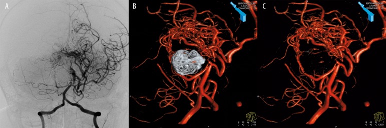
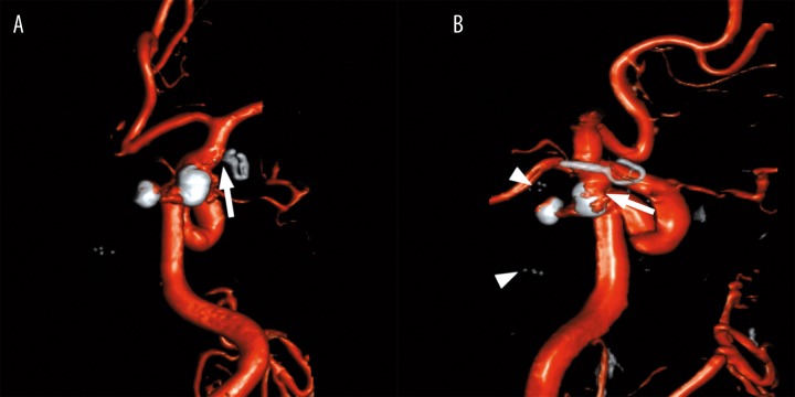
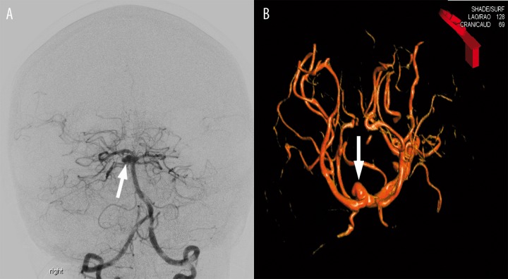
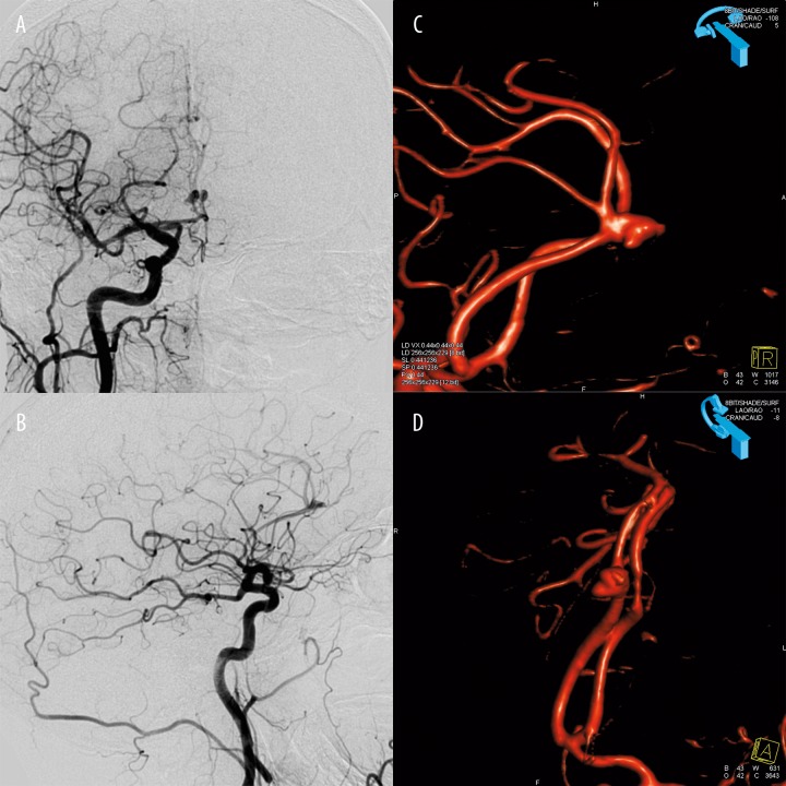
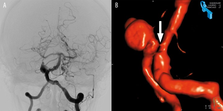
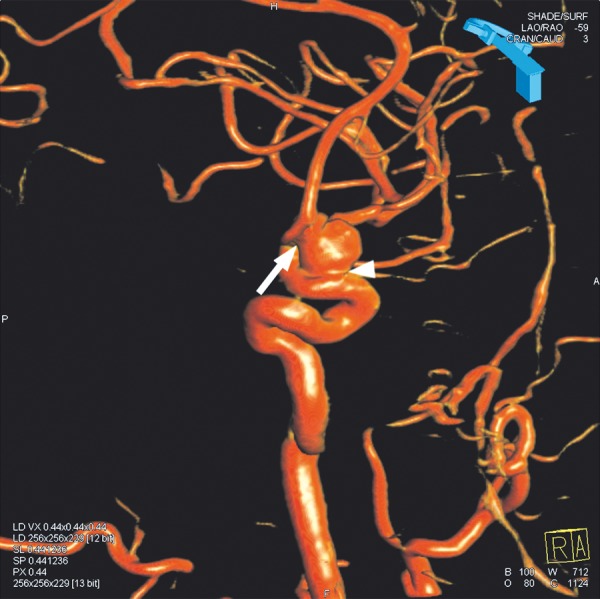
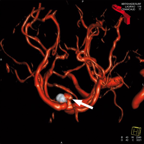
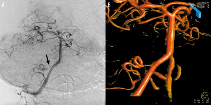
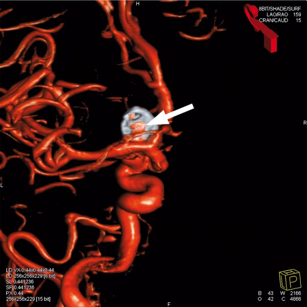
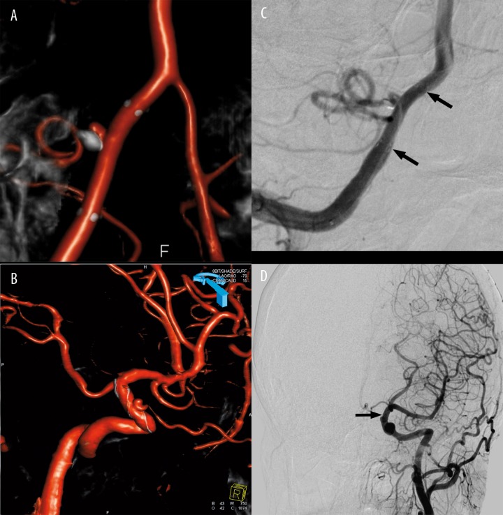
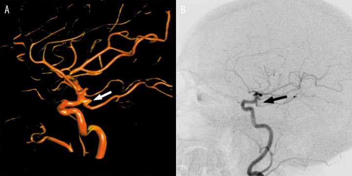
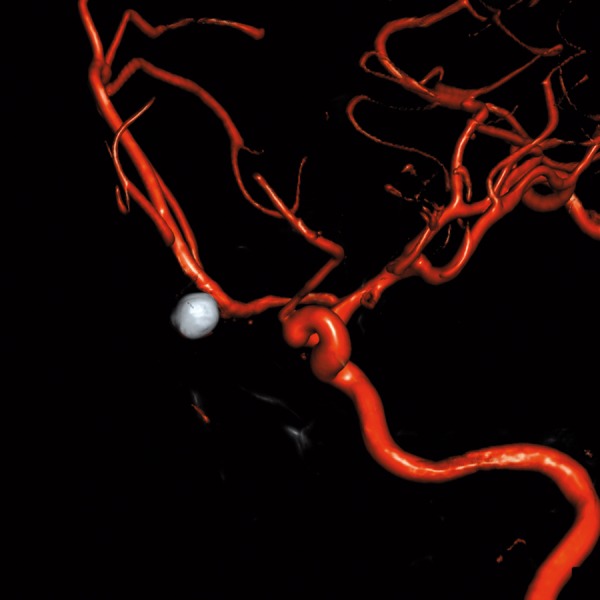
A gold standard of cerebral vessel imaging remains the digital subtraction angiography (DSA) performed in three projections. However, in specific clinical cases, many additional projections are required, or a complete visualization of a lesion may even be impossible with 2D angiography. Three-dimensional (3D) reconstructions of rotational angiography were reported to improve the performance of DSA significantly. In this pictorial essay, specific applications of this technique are presented in the management of intracranial aneurysms, including: preoperative aneurysm evaluation, intraoperative imaging, and follow-up. Volumetric reconstructions of 3D DSA are a valuable tool for cerebral vessels imaging. They play a vital role in the assessment of intracranial aneurysms, especially in evaluation of the aneurysm neck and the aneurysm recanalization.

摘要

脑血管成像的金标准仍然是在三个投影方向上进行的数字减影血管造影(DSA)。然而,在特定临床病例中,需要许多额外的投影方向,甚至二维血管造影可能无法完整显示病变。据报道,旋转血管造影的三维(3D)重建可显著提高DSA的性能。在这篇图文并茂的文章中,介绍了该技术在颅内动脉瘤治疗中的具体应用,包括:术前动脉瘤评估、术中成像和随访。三维DSA的容积重建是脑血管成像的一种有价值的工具。它们在颅内动脉瘤的评估中起着至关重要的作用,尤其是在评估动脉瘤颈部和动脉瘤再通方面。

https://cdn.ncbi.nlm.nih.gov/pmc/blobs/cfbe/3403801/bbfe80f5ec74/poljradiol-77-2-47-g012.jpg
https://cdn.ncbi.nlm.nih.gov/pmc/blobs/cfbe/3403801/114aa53bd8dc/poljradiol-77-2-47-g001.jpg
https://cdn.ncbi.nlm.nih.gov/pmc/blobs/cfbe/3403801/11c1d597e54b/poljradiol-77-2-47-g002.jpg
https://cdn.ncbi.nlm.nih.gov/pmc/blobs/cfbe/3403801/05d7b87c373f/poljradiol-77-2-47-g003.jpg
https://cdn.ncbi.nlm.nih.gov/pmc/blobs/cfbe/3403801/def11c640969/poljradiol-77-2-47-g004.jpg
https://cdn.ncbi.nlm.nih.gov/pmc/blobs/cfbe/3403801/8f50af2f62e7/poljradiol-77-2-47-g005.jpg
https://cdn.ncbi.nlm.nih.gov/pmc/blobs/cfbe/3403801/87d7f6152867/poljradiol-77-2-47-g006.jpg
https://cdn.ncbi.nlm.nih.gov/pmc/blobs/cfbe/3403801/9d81d955a8e4/poljradiol-77-2-47-g007.jpg
https://cdn.ncbi.nlm.nih.gov/pmc/blobs/cfbe/3403801/ee1500152f1f/poljradiol-77-2-47-g008.jpg
https://cdn.ncbi.nlm.nih.gov/pmc/blobs/cfbe/3403801/d35d2f155909/poljradiol-77-2-47-g009.jpg
https://cdn.ncbi.nlm.nih.gov/pmc/blobs/cfbe/3403801/626c04b23285/poljradiol-77-2-47-g010.jpg
https://cdn.ncbi.nlm.nih.gov/pmc/blobs/cfbe/3403801/d8c02c9cc4b0/poljradiol-77-2-47-g011.jpg
https://cdn.ncbi.nlm.nih.gov/pmc/blobs/cfbe/3403801/bbfe80f5ec74/poljradiol-77-2-47-g012.jpg

相似文献

1
DSA volumetric 3D reconstructions of intracranial aneurysms: A pictorial essay.颅内动脉瘤的数字减影血管造影容积三维重建:图文综述。
Pol J Radiol. 2012 Apr;77(2):47-53. doi: 10.12659/pjr.882970.
2
Three-dimensional reconstructed rotational digital subtraction angiography in planning treatment of intracranial aneurysms.三维重建旋转数字减影血管造影术在颅内动脉瘤治疗规划中的应用
Eur Radiol. 2000;10(4):564-8. doi: 10.1007/s003300050961.
3
Evaluation of a distal pericallosal aneurysm visualized with 3-dimensional digital subtraction angiography: case report and treatment implications.三维数字减影血管造影显示的胼胝体远端动脉瘤评估:病例报告及治疗意义
Surg Neurol. 2005 Oct;64(4):321-4. doi: 10.1016/j.surneu.2004.11.027.
4
Comparison of 2D and 3D digital subtraction angiography in evaluation of intracranial aneurysms.二维和三维数字减影血管造影在颅内动脉瘤评估中的比较。
AJNR Am J Neuroradiol. 2002 Oct;23(9):1545-52.
5
Three-dimensional digital subtraction angiography vs two-dimensional digital subtraction angiography for detection of ruptured intracranial aneurysms: a study of 86 aneurysms.三维数字减影血管造影术与二维数字减影血管造影术用于检测破裂颅内动脉瘤的比较:86例动脉瘤的研究
Neurol India. 2005 Sep;53(3):287-9; discussion 290. doi: 10.4103/0028-3886.16925.
6
Dual-energy CT angiography in the evaluation of intracranial aneurysms: image quality, radiation dose, and comparison with 3D rotational digital subtraction angiography.双能量 CT 血管造影在颅内动脉瘤评估中的应用:图像质量、辐射剂量,以及与三维旋转数字减影血管造影的比较。
AJR Am J Roentgenol. 2010 Jan;194(1):23-30. doi: 10.2214/AJR.08.2290.
7
The role of MR angiography in the pretreatment assessment of intracranial aneurysms: a comparative study.磁共振血管造影在颅内动脉瘤术前评估中的作用:一项对比研究。
AJNR Am J Neuroradiol. 2000 Oct;21(9):1618-28.
8
[Digital 3D rotational angiography for the preoperative and preinterventional clarification of cerebral arterial aneurysms].数字3D旋转血管造影术用于脑动脉瘤术前及介入前的评估
Rofo. 1999 May;170(5):482-91. doi: 10.1055/s-2007-1011077.
9
Benefits of 3D rotational DSA compared with 2D DSA in the evaluation of intracranial aneurysm.3D 旋转 DSA 在颅内动脉瘤评估中的优势与 2D DSA 比较。
Acad Radiol. 2012 Jun;19(6):701-7. doi: 10.1016/j.acra.2012.02.012.
10
Comparison of 2D digital subtraction angiography and 3D rotational angiography in the evaluation of dome-to-neck ratio.二维数字减影血管造影与三维旋转血管造影在评估穹顶与颈部比例中的比较。
AJNR Am J Neuroradiol. 2009 Apr;30(4):831-4. doi: 10.3174/ajnr.A1444. Epub 2009 Jan 8.

引用本文的文献

1
Kissing Aneurysms of the Anterior Communicating Artery Treated With Surgical Clipping: A Case Report and Literature Review.手术夹闭治疗前交通动脉的接吻动脉瘤:一例报告及文献复习
Cureus. 2024 May 22;16(5):e60824. doi: 10.7759/cureus.60824. eCollection 2024 May.
2
Self-supervised learning enables 3D digital subtraction angiography reconstruction from ultra-sparse 2D projection views: A multicenter study.基于自监督学习的超稀疏二维投影视图三维数字减影血管造影重建:一项多中心研究。
Cell Rep Med. 2022 Oct 18;3(10):100775. doi: 10.1016/j.xcrm.2022.100775. Epub 2022 Oct 7.
3
Reliability of dual- vs single-volume reconstruction of three-dimensional digital subtraction angiography for follow-up evaluation of endovascularly treated intracranial aneurysms.

本文引用的文献

1
Intracranial aneurysms treated with coil placement: test characteristics of follow-up MR angiography--multicenter study.颅内动脉瘤采用线圈放置治疗:随访磁共振血管造影的测试特征——多中心研究。
Radiology. 2010 Jul;256(1):209-18. doi: 10.1148/radiol.10091528. Epub 2010 May 26.
2
Image quality and artefact generation post-cerebral aneurysm clipping using a 64-row multislice computer tomography angiography (MSCTA) technology: A retrospective study and review of the literature.使用64排多层计算机断层血管造影(MSCTA)技术进行脑动脉瘤夹闭术后的图像质量与伪影产生:一项回顾性研究及文献综述
Clin Neurol Neurosurg. 2010 Jun;112(5):386-91. doi: 10.1016/j.clineuro.2010.02.001. Epub 2010 Mar 1.
3
三维数字减影血管造影双容积与单容积重建在血管内治疗颅内动脉瘤随访评估中的可靠性
Interv Neuroradiol. 2016 Dec;22(6):687-692. doi: 10.1177/1591019916663469. Epub 2016 Aug 16.
Three-dimensional volume-rendering technique in the angiographic follow-up of intracranial aneurysms embolized with coils.
三维容积再现技术在颅内动脉瘤血管内栓塞后血管造影随访中的应用。
J Neurosurg. 2010 Mar;112(3):674-80. doi: 10.3171/2009.7.JNS081718.
4
Opinion: imaging follow-up after coiling of intracranial aneurysms.观点:颅内动脉瘤弹簧圈栓塞后的影像学随访。
AJNR Am J Neuroradiol. 2009 Oct;30(9):1646-8. doi: 10.3174/ajnr.A1673. Epub 2009 Jul 17.
5
Comparison of 2D digital subtraction angiography and 3D rotational angiography in the evaluation of dome-to-neck ratio.二维数字减影血管造影与三维旋转血管造影在评估穹顶与颈部比例中的比较。
AJNR Am J Neuroradiol. 2009 Apr;30(4):831-4. doi: 10.3174/ajnr.A1444. Epub 2009 Jan 8.
6
3D rotational angiography: the new gold standard in the detection of additional intracranial aneurysms.3D旋转血管造影术:检测额外颅内动脉瘤的新金标准。
AJNR Am J Neuroradiol. 2008 May;29(5):976-9. doi: 10.3174/ajnr.A0964. Epub 2008 Feb 7.
7
3D rotational digital subtraction angiography may underestimate intracranial aneurysms: findings from two basilar aneurysms.三维旋转数字减影血管造影术可能低估颅内动脉瘤:两个基底动脉瘤的研究结果
AJNR Am J Neuroradiol. 2007 Oct;28(9):1690-2. doi: 10.3174/ajnr.A0652. Epub 2007 Sep 24.
8
Flat panel detector angiographic CT for stent-assisted coil embolization of broad-based cerebral aneurysms.平板探测器血管造影CT用于宽颈脑动脉瘤的支架辅助弹簧圈栓塞术
AJNR Am J Neuroradiol. 2007 Nov-Dec;28(10):1902-8. doi: 10.3174/ajnr.A0697. Epub 2007 Sep 24.
9
Angiogram-negative subarachnoid hemorrhage in the era of three dimensional rotational angiography.三维旋转血管造影时代的血管造影阴性蛛网膜下腔出血
J Clin Neurosci. 2007 Mar;14(3):252-5. doi: 10.1016/j.jocn.2006.01.011.
10
Comparison of 2D and 3D digital subtraction angiography in evaluation of intracranial aneurysms.二维和三维数字减影血管造影在颅内动脉瘤评估中的比较。
AJNR Am J Neuroradiol. 2002 Oct;23(9):1545-52.