Centre of Ophthalmology and Vision Sciences, IBILI, Faculty of Medicine, University of Coimbra, Coimbra, Portugal.
PLoS One. 2012;7(8):e42428. doi: 10.1371/journal.pone.0042428. Epub 2012 Aug 3.
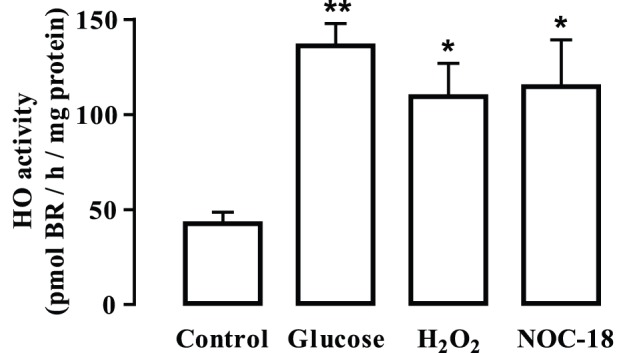
Diabetic retinopathy is a leading cause of visual loss and blindness, characterized by microvascular dysfunction. Hyperglycemia is considered the major pathogenic factor for the development of diabetic retinopathy and is associated with increased oxidative/nitrosative stress in the retina. Since heme oxygenase-1 (HO-1) is an enzyme with antioxidant and protective properties, we investigated the potential protective role of HO-1 in retinal endothelial cells exposed to high glucose and oxidative/nitrosative stress conditions. Retinal endothelial cells were exposed to elevated glucose, nitric oxide (NO) and hydrogen peroxide (H(2)O(2)). Cell viability and apoptosis were assessed by MTT assay, Hoechst staining, TUNEL assay and Annexin V labeling. The production of reactive oxygen species (ROS) was detected by the oxidation of 2',7'-dichlorodihydrofluorescein diacetate. The content of HO-1 was assessed by immunobloting and immunofluorescence. HO activity was determined by bilirubin production. Long-term exposure (7 days) of retinal endothelial cells to elevated glucose decreased cell viability and had no effect on HO-1 content. However, a short-time exposure (24 h) to elevated glucose did not alter cell viability, but increased both the levels of intracellular ROS and HO-1 content. Moreover, the inhibition of HO with SnPPIX unmasked the toxic effect of high glucose and revealed the protection conferred by HO-1. Oxidative/nitrosative stress conditions increased cell death and HO-1 protein levels. These effects of elevated glucose and HO inhibition on cell death were confirmed in primary endothelial cells (HUVECs). When cells were exposed to oxidative/nitrosative stress conditions there was also an increase in retinal endothelial cell death and HO-1 content. The inhibition of HO enhanced ROS production and the toxic effect induced by exposure to H(2)O(2) and NOC-18 (NO donor). Overexpression of HO-1 prevented the toxic effect induced by H(2)O(2) and NOC-18. In conclusion, HO-1 exerts a protective effect in retinal endothelial cells exposed to hyperglycemic and oxidative/nitrosative stress conditions.
糖尿病视网膜病变是视力丧失和失明的主要原因,其特征为微血管功能障碍。高血糖被认为是糖尿病视网膜病变发展的主要致病因素,并且与视网膜中氧化/硝化应激的增加有关。由于血红素加氧酶-1(HO-1)是一种具有抗氧化和保护特性的酶,因此我们研究了 HO-1 在暴露于高葡萄糖和氧化/硝化应激条件下的视网膜内皮细胞中的潜在保护作用。将视网膜内皮细胞暴露于高葡萄糖、一氧化氮(NO)和过氧化氢(H2O2)中。通过 MTT 测定、Hoechst 染色、TUNEL 测定和 Annexin V 标记评估细胞活力和细胞凋亡。通过 2',7'-二氯二氢荧光素二乙酸盐的氧化来检测活性氧物质(ROS)的产生。通过免疫印迹和免疫荧光评估 HO-1 的含量。通过胆红素的产生来确定 HO 活性。将视网膜内皮细胞长期(7 天)暴露于高葡萄糖中会降低细胞活力,而对 HO-1 含量没有影响。然而,短时间(24 小时)暴露于高葡萄糖不会改变细胞活力,但会增加细胞内 ROS 和 HO-1 含量。此外,用 SnPPIX 抑制 HO 会揭示高葡萄糖的毒性作用,并显示出 HO-1 的保护作用。氧化/硝化应激条件增加了细胞死亡和 HO-1 蛋白水平。在原代内皮细胞(HUVECs)中证实了高葡萄糖和 HO 抑制对细胞死亡的这些影响。当细胞暴露于氧化/硝化应激条件时,视网膜内皮细胞的死亡和 HO-1 含量也会增加。抑制 HO 会增加 ROS 的产生并增强暴露于 H2O2 和 NOC-18(NO 供体)引起的毒性作用。HO-1 的过表达可防止 H2O2 和 NOC-18 引起的毒性作用。总之,HO-1 在暴露于高血糖和氧化/硝化应激条件下的视网膜内皮细胞中发挥保护作用。