Child Psychiatry Branch, Brain Imaging Unit, National Institute of Mental Health, National Institutes of Health, 10 Center Drive, MSC 1367, Building 10, Room 4 C110, Bethesda, MD, 20892, USA.
Biol Sex Differ. 2012 Aug 21;3(1):19. doi: 10.1186/2042-6410-3-19.
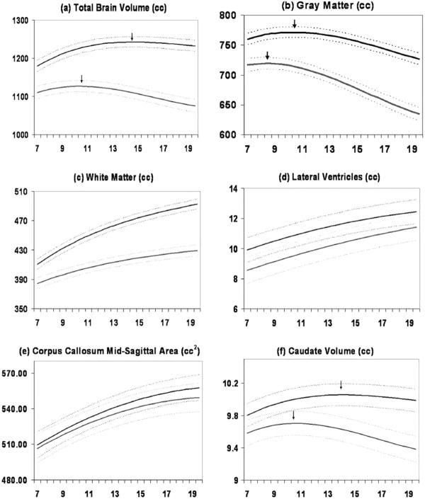
Improvements in neuroimaging technologies, and greater access to their use, have generated a plethora of data regarding male/female differences in the developing brain. Examination of these differences may shed light on the pathophysiology of the many illnesses that differ between the sexes and ultimately lead to more effective interventions. In this review, we attempt to synthesize the anatomic magnetic resonance imaging (MRI) literature of male/female brain differences with emphasis on studies encompassing adolescence - a time of divergence in physical and behavioral characteristics. Across all ages total brain size is consistently reported to be about 10% larger in males. Structures commonly reported to be different between sexes include the caudate nucleus, amygdala, hippocampus, and cerebellum - all noted to have a relatively high density of sex steroid receptors. The direction and magnitude of reported brain differences depends on the methodology of data acquisition and analysis, whether and how the subcomponents are adjusted for the total brain volume difference, and the age of the participants in the studies. Longitudinal studies indicate regional cortical gray matter volumes follow inverted U shaped developmental trajectories with peak size occurring one to three years earlier in females. Cortical gray matter differences are modulated by androgen receptor genotyope and by circulating levels of hormones. White matter volumes increase throughout childhood and adolescence in both sexes but more rapidly in adolescent males resulting in an expanding magnitude of sex differences from childhood to adulthood.
神经影像学技术的进步和更多使用机会的增加,产生了大量关于男性/女性大脑发育差异的数据。研究这些差异可能有助于揭示许多在性别之间存在差异的疾病的病理生理学机制,并最终导致更有效的干预措施。在这篇综述中,我们试图综合男性/女性大脑差异的解剖磁共振成像(MRI)文献,重点是涵盖青春期的研究——这是身体和行为特征差异的时期。在所有年龄段,男性的大脑总体积都被一致报告为比女性大约 10%。通常报告存在性别的结构包括尾状核、杏仁核、海马体和小脑——所有这些结构都被认为具有相对较高密度的性激素受体。报告的脑差异的方向和幅度取决于数据采集和分析的方法,以及是否以及如何根据大脑总体积差异调整子成分,以及研究中参与者的年龄。纵向研究表明,区域皮质灰质体积遵循倒 U 形发育轨迹,女性峰值出现时间比男性早一到三年。皮质灰质差异受雄激素受体基因多态性和循环激素水平的调节。白质体积在儿童期和青春期都在增加,但青春期男性的增加速度更快,导致从儿童期到成年期的性别差异幅度扩大。