• 文献检索
  • 文档翻译
  • 深度研究
  • 学术资讯
  • Suppr Zotero 插件Zotero 插件
  • 邀请有礼
  • 套餐&价格
  • 历史记录
应用&插件
Suppr Zotero 插件Zotero 插件浏览器插件Mac 客户端Windows 客户端微信小程序
定价
高级版会员购买积分包购买API积分包
服务
文献检索文档翻译深度研究API 文档MCP 服务
关于我们
关于 Suppr公司介绍联系我们用户协议隐私条款
关注我们

Suppr 超能文献

核心技术专利:CN118964589B侵权必究
粤ICP备2023148730 号-1Suppr @ 2026

文献检索

告别复杂PubMed语法,用中文像聊天一样搜索,搜遍4000万医学文献。AI智能推荐,让科研检索更轻松。

立即免费搜索

文件翻译

保留排版,准确专业,支持PDF/Word/PPT等文件格式,支持 12+语言互译。

免费翻译文档

深度研究

AI帮你快速写综述,25分钟生成高质量综述,智能提取关键信息,辅助科研写作。

立即免费体验

抗肌萎缩蛋白和优球蛋白素 spectrin 重复的晶体结构:对结构域边界的影响。

The crystal structures of dystrophin and utrophin spectrin repeats: implications for domain boundaries.

机构信息

Institute of Molecular BioSciences, Massey University, Palmerston North, New Zealand.

出版信息

PLoS One. 2012;7(7):e40066. doi: 10.1371/journal.pone.0040066. Epub 2012 Jul 20.

DOI:10.1371/journal.pone.0040066
PMID:22911693
原文链接:https://pmc.ncbi.nlm.nih.gov/articles/PMC3401230/
Abstract

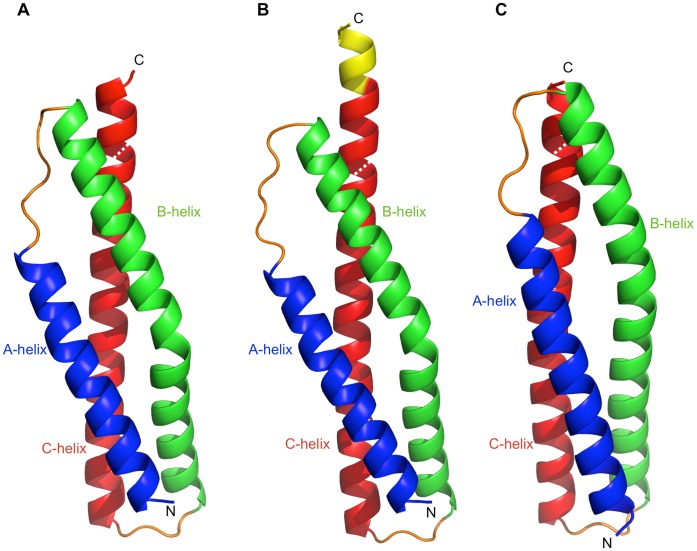
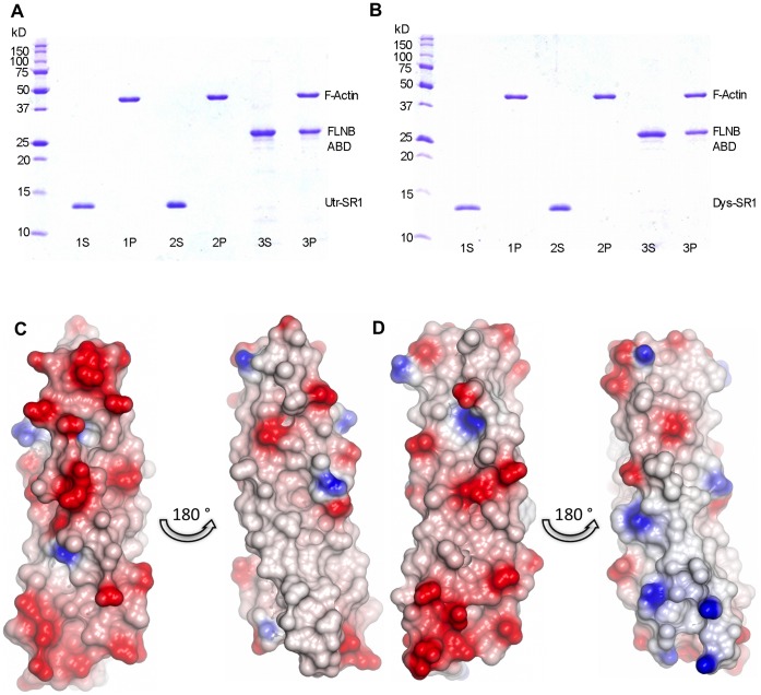
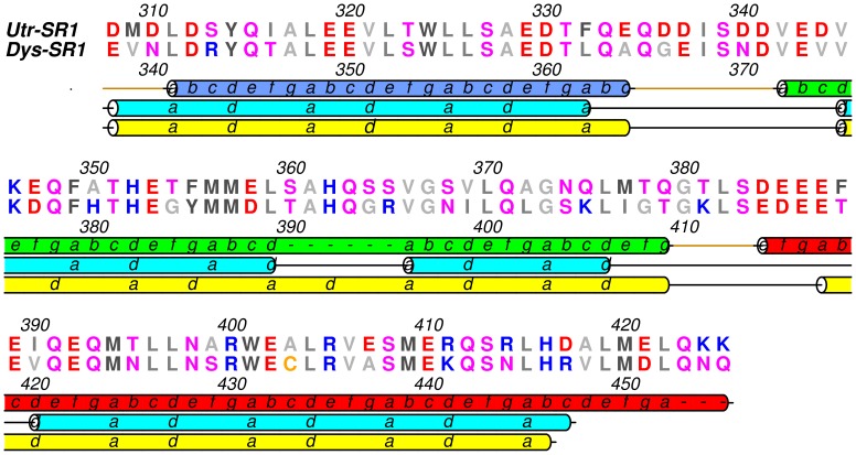
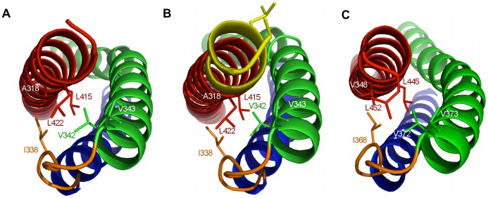
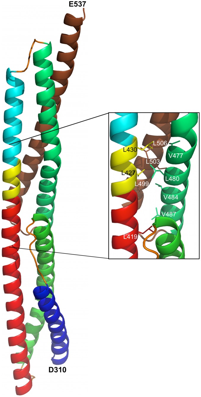
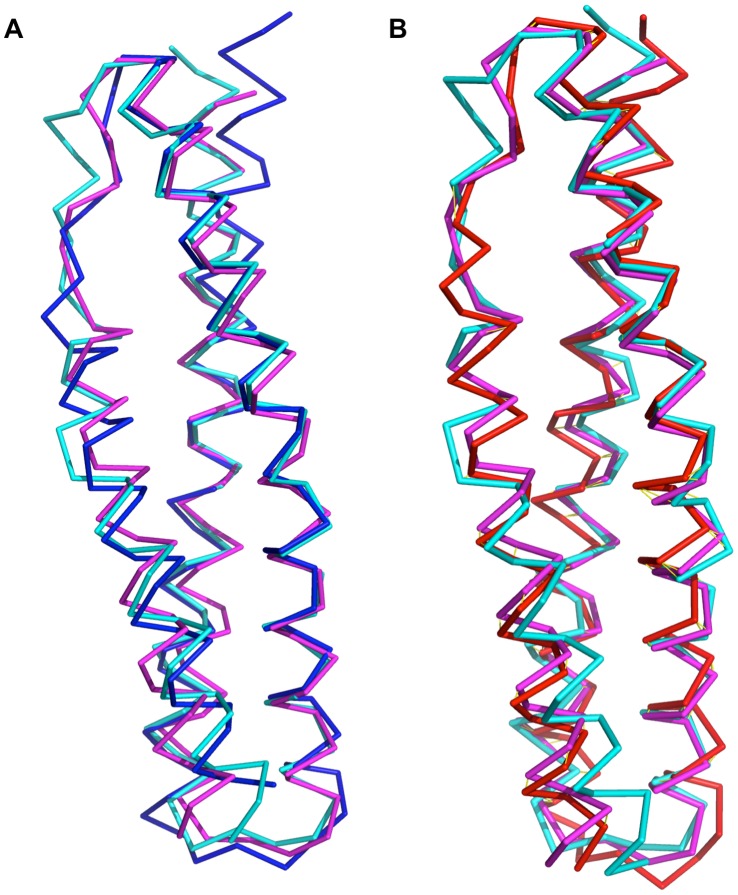
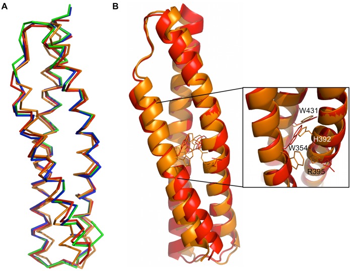
Dystrophin and utrophin link the F-actin cytoskeleton to the cell membrane via an associated glycoprotein complex. This functionality results from their domain organization having an N-terminal actin-binding domain followed by multiple spectrin-repeat domains and then C-terminal protein-binding motifs. Therapeutic strategies to replace defective dystrophin with utrophin in patients with Duchenne muscular dystrophy require full-characterization of both these proteins to assess their degree of structural and functional equivalence. Here the high resolution structures of the first spectrin repeats (N-terminal repeat 1) from both dystrophin and utrophin have been determined by x-ray crystallography. The repeat structures both display a three-helix bundle fold very similar to one another and to homologous domains from spectrin, α-actinin and plectin. The utrophin and dystrophin repeat structures reveal the relationship between the structural domain and the canonical spectrin repeat domain sequence motif, showing the compact structural domain of spectrin repeat one to be extended at the C-terminus relative to its previously defined sequence repeat. These structures explain previous in vitro biochemical studies in which extending dystrophin spectrin repeat domain length leads to increased protein stability. Furthermore we show that the first dystrophin and utrophin spectrin repeats have no affinity for F-actin in the absence of other domains.

摘要

肌营养不良蛋白和 utrophin 通过相关糖蛋白复合物将 F-肌动蛋白细胞骨架连接到细胞膜上。这种功能源自它们的结构域组织,具有 N 端肌动蛋白结合结构域,随后是多个 spectrin 重复结构域,然后是 C 端蛋白结合基序。在杜氏肌营养不良症患者中用 utrophin 替代有缺陷的肌营养不良蛋白的治疗策略需要对这两种蛋白进行充分表征,以评估其结构和功能等效程度。在这里,通过 X 射线晶体学确定了来自肌营养不良蛋白和 utrophin 的第一个 spectrin 重复(N 端重复 1)的高分辨率结构。重复结构均显示出三螺旋束折叠,彼此非常相似,与 spectrin、α-actinin 和 plectin 的同源结构域也非常相似。utrophin 和 dystrophin 重复结构揭示了结构域与规范 spectrin 重复结构域序列基序之间的关系,显示 spectrin 重复 1 的紧凑结构域在 C 末端相对于其先前定义的序列重复得到扩展。这些结构解释了先前的体外生化研究,其中延长肌营养不良蛋白 spectrin 重复结构域的长度会导致蛋白质稳定性增加。此外,我们还表明,在没有其他结构域的情况下,第一个肌营养不良蛋白和 utrophin spectrin 重复结构域与 F-肌动蛋白没有亲和力。

https://cdn.ncbi.nlm.nih.gov/pmc/blobs/e0e6/3401230/490c277cdc90/pone.0040066.g007.jpg
https://cdn.ncbi.nlm.nih.gov/pmc/blobs/e0e6/3401230/d82ceba29a08/pone.0040066.g001.jpg
https://cdn.ncbi.nlm.nih.gov/pmc/blobs/e0e6/3401230/780b4888926b/pone.0040066.g002.jpg
https://cdn.ncbi.nlm.nih.gov/pmc/blobs/e0e6/3401230/bb92053551ea/pone.0040066.g003.jpg
https://cdn.ncbi.nlm.nih.gov/pmc/blobs/e0e6/3401230/35f9f9fc104c/pone.0040066.g004.jpg
https://cdn.ncbi.nlm.nih.gov/pmc/blobs/e0e6/3401230/b6df5b61764b/pone.0040066.g005.jpg
https://cdn.ncbi.nlm.nih.gov/pmc/blobs/e0e6/3401230/a0195b5765ab/pone.0040066.g006.jpg
https://cdn.ncbi.nlm.nih.gov/pmc/blobs/e0e6/3401230/490c277cdc90/pone.0040066.g007.jpg

相似文献

1
The crystal structures of dystrophin and utrophin spectrin repeats: implications for domain boundaries.抗肌萎缩蛋白和优球蛋白素 spectrin 重复的晶体结构:对结构域边界的影响。
PLoS One. 2012;7(7):e40066. doi: 10.1371/journal.pone.0040066. Epub 2012 Jul 20.
2
An atomic model for actin binding by the CH domains and spectrin-repeat modules of utrophin and dystrophin.关于肌动蛋白与肌养蛋白和肌营养不良蛋白的CH结构域及血影蛋白重复模块结合的原子模型。
J Mol Biol. 2003 May 23;329(1):15-33. doi: 10.1016/s0022-2836(03)00422-4.
3
The N- and C-Terminal Domains Differentially Contribute to the Structure and Function of Dystrophin and Utrophin Tandem Calponin-Homology Domains.N 端和 C 端结构域对肌营养不良蛋白和厄普蛋白串联钙调蛋白同源结构域的结构和功能有不同贡献。
Biochemistry. 2015 Nov 24;54(46):6942-50. doi: 10.1021/acs.biochem.5b00969. Epub 2015 Nov 13.
4
The 2.0 A structure of the second calponin homology domain from the actin-binding region of the dystrophin homologue utrophin.来自肌营养不良蛋白同源物Utrophin肌动蛋白结合区域的第二个钙调蛋白同源结构域的2.0埃结构。
J Mol Biol. 1999 Jan 22;285(3):1257-64. doi: 10.1006/jmbi.1998.2406.
5
The structure of the N-terminal actin-binding domain of human dystrophin and how mutations in this domain may cause Duchenne or Becker muscular dystrophy.人类肌营养不良蛋白N端肌动蛋白结合结构域的结构以及该结构域中的突变如何导致杜氏或贝克型肌营养不良症。
Structure. 2000 May 15;8(5):481-91. doi: 10.1016/s0969-2126(00)00132-5.
6
Crystal structure of the actin-binding region of utrophin reveals a head-to-tail dimer.肌动蛋白结合蛋白肌养蛋白的肌动蛋白结合区域的晶体结构揭示了一种头对头二聚体。
Structure. 1999 Dec 15;7(12):1539-46. doi: 10.1016/s0969-2126(00)88344-6.
7
Dystrophin and utrophin bind actin through distinct modes of contact.肌营养不良蛋白和抗肌萎缩蛋白通过不同的接触方式结合肌动蛋白。
J Biol Chem. 2006 Apr 14;281(15):9996-10001. doi: 10.1074/jbc.M513121200. Epub 2006 Feb 13.
8
Interdomain Linker Determines Primarily the Structural Stability of Dystrophin and Utrophin Tandem Calponin-Homology Domains Rather than Their Actin-Binding Affinity.结构域间连接子主要决定肌营养不良蛋白和肌萎缩蛋白串联钙调蛋白同源结构域的结构稳定性,而非其肌动蛋白结合亲和力。
Biochemistry. 2015 Sep 8;54(35):5480-8. doi: 10.1021/acs.biochem.5b00741. Epub 2015 Aug 26.
9
Distinct mechanical properties in homologous spectrin-like repeats of utrophin.肌萎缩蛋白同源 spectrin 样重复区具有独特的力学性能。
Sci Rep. 2019 Mar 26;9(1):5210. doi: 10.1038/s41598-019-41569-4.
10
The 8th and 9th tandem spectrin-like repeats of utrophin cooperatively form a functional unit to interact with polarity-regulating kinase PAR-1b.肌萎缩蛋白的第 8 和第 9 串联 spectrin 样重复结构域共同形成一个功能单位,与极性调节激酶 PAR-1b 相互作用。
Biochem Biophys Res Commun. 2010 Jan 1;391(1):812-7. doi: 10.1016/j.bbrc.2009.11.144. Epub 2009 Nov 27.

引用本文的文献

1
Structure and assembly of the dystrophin glycoprotein complex.肌营养不良蛋白糖蛋白复合体的结构与组装
Nature. 2025 Jan;637(8048):1252-1260. doi: 10.1038/s41586-024-08310-2. Epub 2024 Dec 11.
2
Coupling of Spectrin Repeat Modules for the Assembly of Nanorods and Presentation of Protein Domains. spectrin 重复模块的偶联对于纳米棒的组装和蛋白质结构域的呈现。
ACS Nano. 2024 Oct 22;18(42):28748-28763. doi: 10.1021/acsnano.4c07701. Epub 2024 Oct 11.
3
Combining Protein Expression and Molecular Data Improves Mutation Characterization of Dystrophinopathies.

本文引用的文献

1
Internal deletion compromises the stability of dystrophin.内部缺失会损害肌营养不良蛋白的稳定性。
Hum Mol Genet. 2011 Aug 1;20(15):2955-63. doi: 10.1093/hmg/ddr199. Epub 2011 May 10.
2
Crystal structure of a rigid four-spectrin-repeat fragment of the human desmoplakin plakin domain.人桥蛋白 plakinin 结构域刚性四 spectrin 重复片段的晶体结构。
J Mol Biol. 2011 Jun 24;409(5):800-12. doi: 10.1016/j.jmb.2011.04.046. Epub 2011 Apr 22.
3
ConSurf 2010: calculating evolutionary conservation in sequence and structure of proteins and nucleic acids.
结合蛋白质表达和分子数据可改善肌营养不良症的突变特征分析。
Front Neurol. 2021 Dec 7;12:718396. doi: 10.3389/fneur.2021.718396. eCollection 2021.
4
Mechanical Unfolding of Spectrin Repeats Induces Water-Molecule Ordering.机械展开 spectrin 重复序列诱导水分子有序排列。
Biophys J. 2020 Mar 10;118(5):1076-1089. doi: 10.1016/j.bpj.2020.01.005. Epub 2020 Jan 16.
5
Empirical and Computational Comparison of Alternative Therapeutic Exon Skip Repairs for Duchenne Muscular Dystrophy.替代治疗外显子跳跃修复杜氏肌营养不良症的实验和计算比较。
Biochemistry. 2019 Apr 16;58(15):2061-2076. doi: 10.1021/acs.biochem.9b00062. Epub 2019 Apr 1.
6
Development of Novel Micro-dystrophins with Enhanced Functionality.新型功能增强型微肌营养不良蛋白的研发。
Mol Ther. 2019 Mar 6;27(3):623-635. doi: 10.1016/j.ymthe.2019.01.002. Epub 2019 Feb 1.
7
Syntrophins entangled in cytoskeletal meshwork: Helping to hold it all together.与细胞骨架网纠缠在一起的连接蛋白:帮助将其全部固定在一起。
Cell Prolif. 2019 Mar;52(2):e12562. doi: 10.1111/cpr.12562. Epub 2018 Dec 4.
8
Fine mapping of hydrophobic contacts reassesses the organization of the first three dystrophin coiled-coil repeats.精确定位疏水区接触点,重新评估前三个肌营养不良蛋白卷曲螺旋重复的结构。
Protein Sci. 2019 Mar;28(3):561-570. doi: 10.1002/pro.3557. Epub 2019 Jan 14.
9
Syntrophin binds directly to multiple spectrin-like repeats in dystrophin and mediates binding of nNOS to repeats 16-17.联蛋白直接与 dystrophin 中的多个 spectrin 样重复序列结合,并介导 nNOS 与重复序列 16-17 的结合。
Hum Mol Genet. 2018 Sep 1;27(17):2978-2985. doi: 10.1093/hmg/ddy197.
10
Mechanical unfolding reveals stable 3-helix intermediates in talin and α-catenin.机械展开揭示了 talin 和 α-连环蛋白中的稳定 3 螺旋中间体。
PLoS Comput Biol. 2018 Apr 26;14(4):e1006126. doi: 10.1371/journal.pcbi.1006126. eCollection 2018 Apr.
ConSurf 2010:计算蛋白质和核酸序列及结构的进化保守性。
Nucleic Acids Res. 2010 Jul;38(Web Server issue):W529-33. doi: 10.1093/nar/gkq399. Epub 2010 May 16.
4
A biophysical map of the dystrophin rod.肌营养不良蛋白杆状区的生物物理图谱。
Biochim Biophys Acta. 2010 Sep;1804(9):1796-809. doi: 10.1016/j.bbapap.2010.03.009. Epub 2010 Apr 9.
5
I-TASSER: a unified platform for automated protein structure and function prediction.I-TASSER:一个用于自动化蛋白质结构和功能预测的统一平台。
Nat Protoc. 2010 Apr;5(4):725-38. doi: 10.1038/nprot.2010.5. Epub 2010 Mar 25.
6
The artificial gene Jazz, a transcriptional regulator of utrophin, corrects the dystrophic pathology in mdx mice.人工基因 Jazz 是肌萎缩蛋白的转录调节剂,可纠正 mdx 小鼠的肌肉营养不良病理。
Hum Mol Genet. 2010 Mar 1;19(5):752-60. doi: 10.1093/hmg/ddp539. Epub 2009 Dec 4.
7
The 8th and 9th tandem spectrin-like repeats of utrophin cooperatively form a functional unit to interact with polarity-regulating kinase PAR-1b.肌萎缩蛋白的第 8 和第 9 串联 spectrin 样重复结构域共同形成一个功能单位,与极性调节激酶 PAR-1b 相互作用。
Biochem Biophys Res Commun. 2010 Jan 1;391(1):812-7. doi: 10.1016/j.bbrc.2009.11.144. Epub 2009 Nov 27.
8
Skeletal dysplasias due to filamin A mutations result from a gain-of-function mechanism distinct from allelic neurological disorders.由于细丝蛋白 A 突变导致的骨骼发育不良是一种不同于等位基因神经紊乱的获得性功能机制。
Hum Mol Genet. 2009 Dec 15;18(24):4791-800. doi: 10.1093/hmg/ddp442. Epub 2009 Sep 22.
9
Pharmacological activation of PPARbeta/delta stimulates utrophin A expression in skeletal muscle fibers and restores sarcolemmal integrity in mature mdx mice.PPARβ/δ 的药理学激活可刺激骨骼肌纤维中的 utrophin A 表达,并恢复成熟 mdx 小鼠的肌细胞膜完整性。
Hum Mol Genet. 2009 Dec 1;18(23):4640-9. doi: 10.1093/hmg/ddp431. Epub 2009 Sep 10.
10
Disease-associated substitutions in the filamin B actin binding domain confer enhanced actin binding affinity in the absence of major structural disturbance: Insights from the crystal structures of filamin B actin binding domains.细丝蛋白B肌动蛋白结合结构域中与疾病相关的替代在无主要结构紊乱的情况下赋予增强的肌动蛋白结合亲和力:来自细丝蛋白B肌动蛋白结合结构域晶体结构的见解
J Mol Biol. 2009 Jul 31;390(5):1030-47. doi: 10.1016/j.jmb.2009.06.009. Epub 2009 Jun 6.